
Rat Intrahepatic Bile Duct Epithelial Cells
Cat.No.: CSC-C5086S
Species: Rat
Source: Bile Duct
Cell Type: Epithelial Cell; Cholangiocyte
- Specification
- Background
- Scientific Data
- Q & A
- Customer Review
Rat Intrahepatic Bile Duct Epithelial Cells (rIBDECs) from Creative Bioarray are isolated from the rat liver tissue. The method we use to isolate rIBDECs was developed based on a combination of established and our proprietary methods. The rIBDECs are characterized by immunofluorescence with antibodies specific to cytokeratin-19 (CK-19). Each vial contains 0.5x10^6 cells per ml and is delivered frozen.
Intrahepatic bile duct epithelial cells (IBDECs), also termed cholangiocytes, constitute a specialized epithelial population lining the biliary tree within the liver. Though comprising only 3–5% of hepatic cells, they are essential for modifying hepatocyte-derived canalicular bile through tightly regulated secretory and absorptive processes, as well as participating in liver injury responses, inflammation, and fibrogenesis. Primary rat IBDECs, isolated via collagenase perfusion and immunomagnetic purification, provide an exceptionally faithful in vitro system that captures native cholangiocyte biology.
A principal strength of primary rat IBDECs is the retention of differentiated epithelial features. They form confluent, polarized monolayers with functional tight junctions and exhibit strong expression of cholangiocyte markers, including cytokeratin 19, γ-glutamyltransferase, and aquaporin 1. Functional secretin receptors are preserved, eliciting cAMP-dependent CFTR activation and vectorial chloride and bicarbonate secretion, thereby recapitulating ductal bile formation. The cells also bear primary cilia that serve as mechano- and chemosensory organelles, and they respond robustly to growth factors (VEGF, HGF) and pro-inflammatory cytokines, enabling mechanistic studies of biliary proliferation, apoptosis, senescence, and epithelial-mesenchymal transition.
Compared with immortalized cholangiocyte lines, primary rat IBDECs avoid transformed phenotypes and genomic instability, preserving native transporter, ion channel, and receptor profiles, as well as physiological hormone responsiveness. The rat origin confers practical advantages: high cell yields from a single liver, lower cost and fewer ethical constraints than human tissue, and seamless integration with established rat models of cholestasis (bile duct ligation, ANIT-induced) and fibrosis. This concordance allows direct in vitro–in vivo correlation of molecular findings.
The Clostridium Metabolite P-Cresol Sulfate Relieves Inflammation of Primary Biliary Cholangitis by Regulating Kupffer Cells
Current study aimed to explore the effects and mechanisms of the Clostridium metabolite p-Cresol sulfate (PCS) in primary biliary cholangitis (PBC).
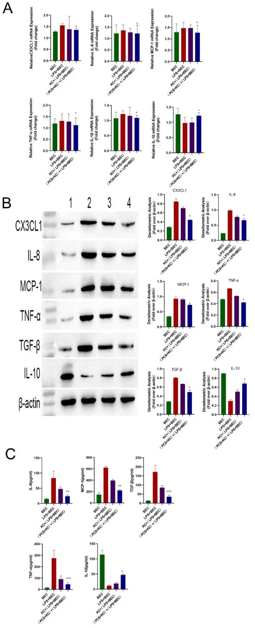
Previous studies found that in PBC mice, Kupffer cells are activated and damage bile duct epithelial cells via cytokines and participate in the development of diseases. To determine whether PCS can protect damaged bile duct epithelial cells via Kupffer cells, we administered LPS to damage bile duct epithelial cells, followed by their co-culture with PCS-stimulated Kupffer cells. Co-cultures were divided into four groups: bile duct epithelial cells cultured alone (BEC); 100 ng/mL LPS administered to bile duct epithelial cells and cultured for 4 h (BEC + LPS); co-culture of Kupffer cells with LPS-injured bile duct epithelial cells for 12 h KC + (BEC + LPS); and Kupffer cells treated with 200 μg/mL PCS prior to co-culture with LPS-injured bile duct epithelial cells for 12 h (PCS + KC) + (BEC + LPS).
Following LPS stimulation, we observed elevated mRNA levels of inflammatory cytokines (TNF-α, IL-8, MCP-1, IL-1β, and CX3CL1) and decreased mRNA levels of the anti-inflammatory cytokine IL-10 relative to levels observed in the BEC group and consistent with previous results and a clinical PBC phenotype. However, in the (PCS + KC) + (BEC + LPS) group, both mRNA and protein levels of these cytokines recovered to those observed in the BEC group (Fig. 1A, B). Kupffer cells without PCS also have the effect of reducing inflammation, but the effect was expanded by PCS. These results were subsequently confirmed by ELISA (Fig. 1C), suggesting that PCS-stimulated Kupffer cells protected LPS-damaged bile duct epithelial cells.
Ask a Question
Write your own review
- You May Also Need
Description: The thoracic aorta is located in the chest cavity and gives off arteries that branch to the esophagus, pericardium, lungs, and trachea. The thoracic aorta can be subdivided into the ascending aorta, ...
Description: Rat Podocytes are isolated from normal rat kidney. The cells are characterized by immunofluorescence with antibodies specific to podocin, Ang1, Nephrin, ACTN4, NPHS2. T25 flasks is required for cell ...
Description: Rat Bronchial Smooth Muscle Cells are isolated from normal rat bronchi tissue. Rat Bronchial Smooth Muscle Cells are characterized by immunofluorescence with antibodies specific to alpha-actin. T25 ...
Description: Guinea Pig Endothelial Cells from Creative Bioarray are isolated from guinea pig tissue. Prior to shipping, cells at passage 2 are detached from flasks and immediately cryopreserved in vials. Each ...
Description: Rat Lung Epithelial Cells are isolated from normal rat lung tissue. The cells are characterized by immunofluorescence with antibodies specific to CK-18, CK-19. T25 flasks is required for cell ...
Description: Rat Vein Endothelial Cells from Creative Bioarray are isolated from inferior vena cava tissue of 6-8 week old laboratory Sprague-Dawley rat. Rat Vein Endothelial Cells are grown in T75 tissue culture ...