
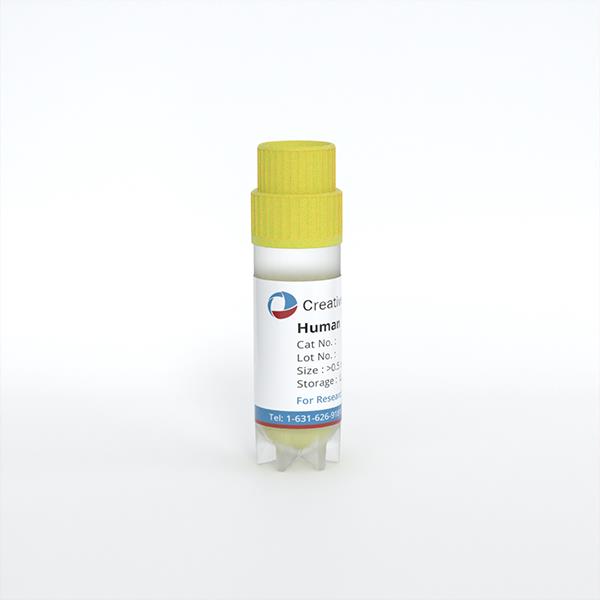
Human Dermal Lymphatic Endothelial Cells
Cat.No.: CSC-C8596W
Species: Human
Source: Dermis; Skin
Cell Type: Endothelial Cell
- Specification
- Background
- Scientific Data
- Q & A
- Customer Review
Human dermal lymphatic endothelial cells (HDLECs) are a type of monolayer flat epithelium that are tightly arranged on the inner surface of lymphatic vessels, forming the primary structure of the lymphatic vessel wall. Through their precise arrangement and structure, they ensure effective lymph transportation and the normal functioning of the lymphatic system. HDLECs typically lack a complete basement membrane, which endows them with high permeability, favoring the exchange of substances between lymphatic fluid and tissue fluid. Moreover, HDLECs possess numerous invaginations and cytoplasmic vesicles, along with characteristic overlapping intercellular junctions. These structural features enhance the strength of intercellular connections and help maintain the integrity and stability of lymphatic vessels. Additionally, HDLECs exhibit positive reactions to specific immunofluorescence markers such as CD31, Podoplanin, and Lyve1, which are commonly used for the identification and isolation of these cells.
HDLECs play a vital role in maintaining physiological balance and immune functionality within the human body. They not only manage fluid, protein and tissue pressure but also serve as sites for lymphocyte recirculation and immune activity. Lymphocytes travel efficiently throughout the body, performing immune functions such as infection prevention and tumour surveillance via lymphatic vessels. Moreover, HDLECs are involved in various pathologic processes including wound healing, lymphoedema, and spreading of inflammation. Several new studies have shown that HDLECs play a pivotal role in tumor metastasis, modulating the spread routes of cancer cells through control of the creation and function of lymphatic vessels In research, HDLECs are often used in making models of lymphatic diseases, including lymphangioma, lymphangitis and lymphatic tuberculosis, in order to understand how these conditions can be treated. Besides these, HDLECs are widely applied to determine how lymphatic endothelial cells multiply, migrate and differentiate, and the molecular signalling networks controlling those functions.
 Fig. 1. Human dermal lymphatic endothelial cells. DAPI: blue nuclei. Corresponding bright field images of CD31 and podoplanin immuno-stained cells appear to the right of each immuno-stained image. (Kong AM, Lim SY, et al., 2022).
Stearic Acid Selectively Induced Apoptosis in Lymphatic Endothelial Cells Through ER Stress and Reactive Oxygen Species
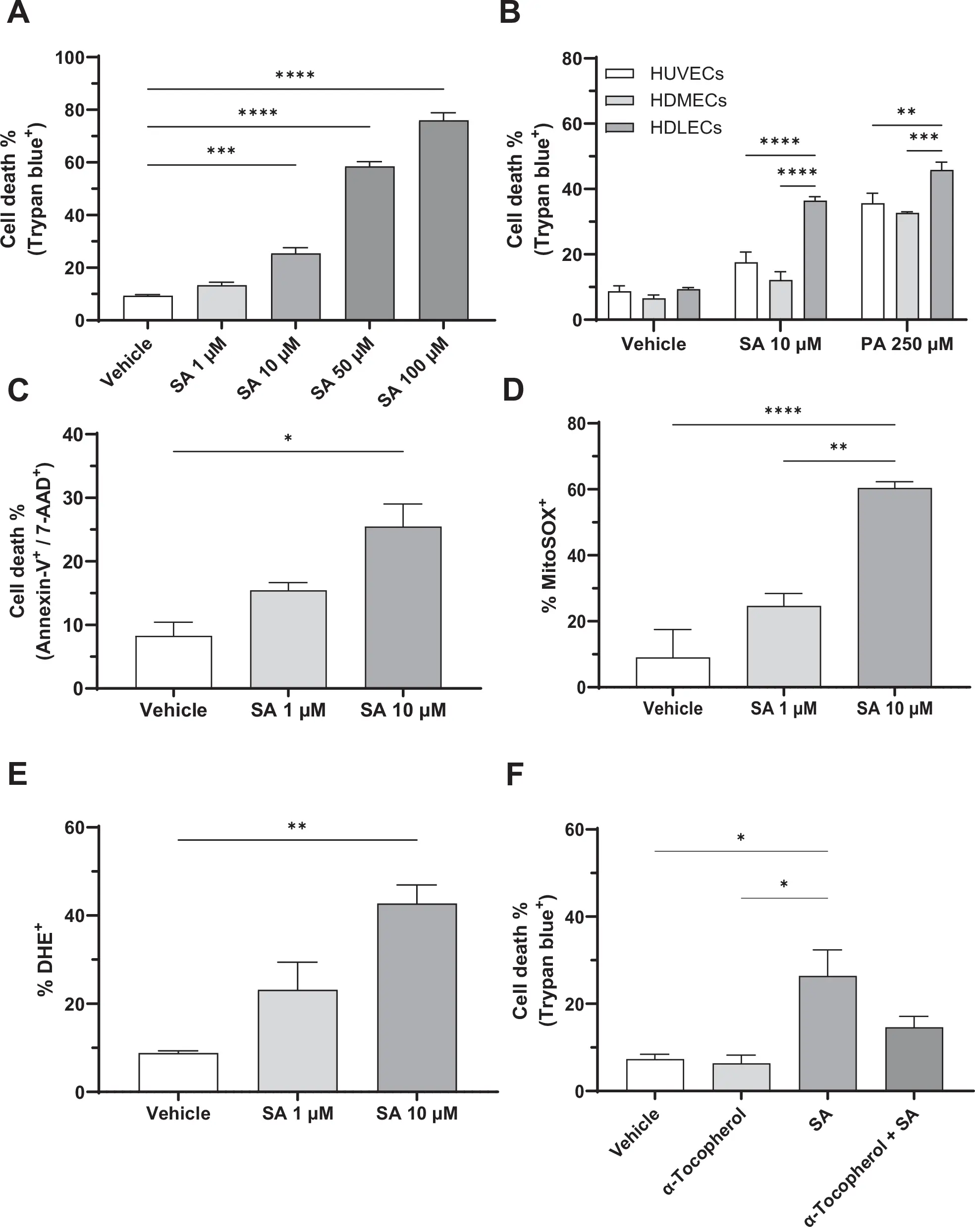
To investigate the in vitro effects of saturated fatty acids (SFAs) on lymphatic and vascular endothelial cells, human dermal lymphatic endothelial cells (HDLECs), human umbilical vein endothelial cells (HUVECs), and human dermal microvascular endothelial cells (HDMECs) were exposed to increasing concentrations of several different fatty acids, and cell death was evaluated using a trypan blue exclusion assay.
Increasing concentrations of the stearic acid (SA) and palmitic acid (PA) resulted in dose-dependent cell death in HDLECs, to a greater extent than HUVECs and HDMECs, indicating that HDLECs are more susceptible to lipotoxicity (Fig. 1A,B,C). These findings suggest that LECs are more susceptible to SFA-induced lipotoxicity than vascular endothelial cells. SA treatment significantly increased reactive oxygen species (ROS) production in HDLECs, with mitochondrial ROS levels elevated by sixfold and intracellular ROS levels elevated by fivefold (Fig. 1D,E,F). In addition, western blot analysis revealed a dose-dependent increase in sXBP-1 and CHOP expression following SA treatment, suggesting that ER stress also contributes to SA-induced cell death in LECs. These findings demonstrate that ROS and ER stress contribute to SA-induced cell death in LECs.
Ask a Question
Write your own review
Description: RFP Expressing Human Dermal Lymphatic Microvascular Endothelial Cells (RFP-HDLMvECs) provided by Creative Bioarray are are puromycin-selected from Human Dermal Lymphatic Microvascular Endothelial ...
Description: Creative Bioarray's normal Human Dermal Fibroblasts-Neonatal, Xeno-Free (HDFn-XF), when grown in Creative Bioarray's LIFib Xeno-Free Complete Medium, provide an ideal xeno-free culture system to ...
Description: Neonatal Human Dermal Fibroblasts (NHDF) are selected by first isolating fibroblasts from the dermis of neonatal skin and then establishing a primary fibroblast culture. Upon completion of the ...
Description: Fibroblasts are mesenchymal cells derived from the embryonic mesoderm. They have been extensively used for a wide range of cellular and molecular studies. This is mainly because they are one of ...
Description: GFP and Luciferase Expressing Human Dermal Fibroblasts (Neonatal) (GFP-Luc-HNDFCs-Neo) provided by Creative Bioarray are puromycin-selected from Human Dermal Fibroblasts infected with lentivirus ...
Description: GFP Expressing Human Dermal Fibroblasts (neonatal) (GFP-HDFCs-Neo) provided by Creative Bioarray are puromycin-selected from Human Dermal Fibroblasts infected with lentivirus expressing GFP. ...