
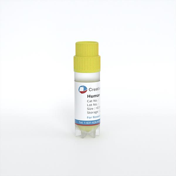
Human Dermal Fibroblasts-fetal-mitomycin C treated (HDF-f-mt)
Cat.No.: CSC-7796W
Species: Human
Source: Dermis; Skin
Cell Type: Fibroblast
- Specification
- Background
- Scientific Data
- Q & A
- Customer Review
Human Dermal Fibroblasts-fetal-mitomycin C treated (HDF-f-mt) are primary fetal human dermal fibroblasts treated with mitomycin C to irreversibly inhibit cell proliferation while maintaining metabolic activity and ability to secrete extracellular matrix components, growth factors and cytokines. These cells can be treated with mitomycin C to make them viable and functional, but incapable of dividing. These cells provide a perfect feeder layer for the expansion and maintenance of diverse epithelial and stem cell populations.
HDF-f-mt cells secrete critical trophic factors and extracellular matrix proteins that create a milieu for cell attachment, survival, proliferation, and differentiation. Widely utilized in keratinocyte culture, growth of epidermal stem cells, skin tissue engineering and regenerative medicine applications. Compared with murine feeder cells, human-derived feeder fibroblasts offer improved physiological relevance and reduce concerns associated with xenogeneic components in translational and clinical research.
These growth-arrested fibroblasts are very useful for the culture of primary human keratinocytes, epithelial progenitor cells, and other fragile cell types that need stromal support. HDF-f-mt cells are also used for experiments in wound healing, artificial skin construction, cell therapies manufacturing and advanced tissue-engineered models. Because they are stable and derived from human cells, they are crucial tools in fundamental research, drug development, and regenerative medicine processes.
Platelet-Rich Fibrin Rescues Mitomycin-C–Induced Fibroblast Senescence with Optimal Effect at 50% Concentration
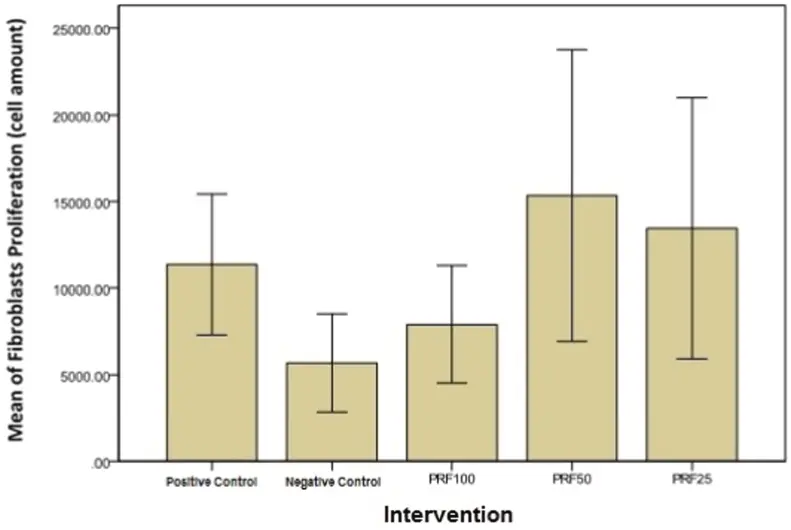
Chronic wounds are characterized by fibroblast senescence and impaired proliferation. Mitomycin-C (MMC) mimics this phenotype by blocking proliferation. Dachlan et al. investigated whether platelet-rich fibrin (PRF) could rescue fibroblast viability after MMC-induced senescence.
MMC significantly reduced fibroblast viability compared to control (p< 0.05) (Fig. 1). PRF supplementation (100%, 50%, 25%) dose-dependently increased viable cell numbers versus MMC alone. Notably, 50% PRF produced the maximal proliferative rescue—surpassing both 25% and undiluted 100% PRF (p< 0.05 among PRF groups). This optimum is attributed to balanced growth factor bioavailability with adequate culture medium nutrients (DMEM + 1% FBS) in diluted PRF, whereas 100% PRF lacked supplemental medium.
These findings align with prior reports that PRF outperforms platelet-rich plasma (PRP) in promoting proliferation, likely due to its higher growth factor content and fibrin scaffold. The dose–response pattern suggests that optimized PRF dilution enhances fibroblast recovery in a senescent-like model, informing future in vivo studies on acute and chronic wound treatment.
Ask a Question
Write your own review
Description: RFP Expressing Human Dermal Lymphatic Microvascular Endothelial Cells (RFP-HDLMvECs) provided by Creative Bioarray are are puromycin-selected from Human Dermal Lymphatic Microvascular Endothelial ...
Description: The melanocyte is a neural crest-derived cell that localizes in humans to several organs including the epidermis, eye, inner ear and leptomeninges. The failure of melanocytes to migrate to these ...
Description: The epithelial layer of the skin provides an essential function as a protective barrier against insult from the outside environment. The major cell type in this layer is keratinocytes which consists ...
Description: Creative Bioarray's normal Human Dermal Fibroblasts-Neonatal, Xeno-Free (HDFn-XF), when grown in Creative Bioarray's LIFib Xeno-Free Complete Medium, provide an ideal xeno-free culture system to ...
Description: Neonatal Human Dermal Fibroblasts (NHDF) are selected by first isolating fibroblasts from the dermis of neonatal skin and then establishing a primary fibroblast culture. Upon completion of the ...
Description: The melanocyte is a neural crest-derived cell that localizes in humans to several organs including the epidermis, eye, inner ear and leptomeninges. The failure of melanocytes to migrate to these ...