
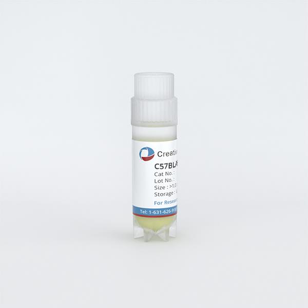
C57BL/6 Mouse Primary Embryonic Fibroblasts
Cat.No.: CSC-C4269X
Species: Mouse
Source: Embryo
Cell Type: Fibroblast
- Specification
- Background
- Scientific Data
- Q & A
- Customer Review
Mouse Primary Embryonic Fibroblasts are negative for bacteria, yeast, fungi, and mycoplasma. Cells are tested for expression of marker using the antibody of anti-FSP1/S100A4 by immunofluorescence staining. Cells can be expanded for 3-6 passages at a split ratio of 1:2 under the cell culture conditions specified by Creative Bioarray. Repeated freezing and thawing of cells is not recommended.Standard biochemical procedures performed with cell cultures include the assay of cell to cell interaction, RT-PCR, Western blotting, immunoprecipitation, immunofluorescent staining, flow cytometry or generating cell derivatives for desired research applications.
Primary embryonic fibroblasts derived from the C57BL/6 mouse strain represent a cornerstone cellular model in modern biomedical research. These cells are isolated from the mesenchyme of trunk, limb buds, and head of pathogen-free embryonic day 13 (E13) mouse embryos, and exhibit the characteristic spindle-shaped morphology of fibroblasts.
The principal advantage of C57BL/6 MEFs lies in their robust proliferative capacity and amenability to isolation and rapid expansion in culture. This inherent durability renders them highly tractable for diverse experimental manipulations, ranging from gene transfection to microinjection. Furthermore, their genetic background is well-characterized, making them a standard model for the functional characterization of gene knockouts, where they provide a defined and reproducible system for studying loss-of-function phenotypes.
Beyond genetic studies, C57BL/6 MEFs are invaluable for their role in stem cell biology. They are widely utilized as feeder layers to support the growth and maintain the undifferentiated, pluripotent state of embryonic stem (ES) and induced pluripotent stem (iPS) cells. This function is mediated by the secretion of critical growth factors and the provision of essential extracellular matrix (ECM) components for cellular adhesion. Their utility extends to disease modeling, serving as an in vitro system to study the pathogenesis of premature aging disorders such as Hutchinson-Gilford progeria, and to investigate mechanisms of cellular reprogramming and genomic instability.
Early-Stage Centrosome Amplification in Primary Mouse Embryonic Fibroblasts
The centrosome is involved in cytoplasmic microtubule organization during interphase and in mitotic spindle assembly during cell division. Centrosome amplification (abnormal proliferation of centrosome number) has been observed in several types of cancer and in precancerous conditions. Therefore, it is important to elucidate the mechanism of centrosome amplification in order to understand the early stage of carcinogenesis.
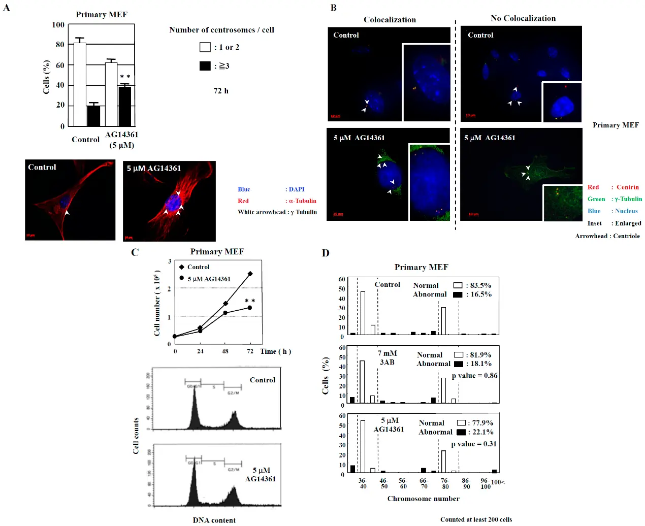
Primary cells could be used to better understand the early stage of carcinogenesis rather than immortalized cells, which tend to have various genetic and epigenetic changes. When primary mouse embryonic fibroblasts (MEF) were incubated with 5 µM AG14361 (a PARP inhibitor known to induce centrosome amplification and chromosomal aneuploidy) for 72 h, there was a significant increase in the number of cells containing three or more spots of γ-tubulin, a marker of the centrosome (Fig. 1A). Moreover, the majority of spots of γ-tubulin colocalized with the spots of centriole, thus confirming that the actual number of centrosomes in primary MEF was increased via AG14361 treatment (Fig. 1B). The proliferation of cells was significantly reduced at 72 h after incubation with 5 µM AG14361 without significant changes in the flow cytometric patterns (Fig. 1C).
In addition to the numerical amplification of centrosomes (centrosome amplification), cancer cells often exhibit changes in the number of chromosomes (aneuploidy) and multipolar spindle formation. In primary MEF, the normal chromosome number was 2n = 40 ± 4 and 80 ± 4 (Fig. 1D). However, aneuploidy was not observed with 5 µM AG14361 treatment in primary MEF (Fig. 1D).
Ask a Question
Write your own review
Description: C57BL/6-GFP Mouse Skeletal Muscle Microvascular Endothelial Cells from Creative Bioarray are isolated from C57BL/6-Tg (CAG-EGFP) 1Osb/J mouse skeletal muscle tissue of pathogen-free laboratory mice. ...
Description: eNOS KO Mouse Stomach Epithelial Cells from Creative Bioarray are isolated from stomach tissue of pathogen-free laboratory mice. eNOS KO Mouse Stomach Epithelial Cells are grown in a T25 tissue ...
Description: eNOS KO Mouse Liver Fibroblasts from Creative Bioarray are isolated from liver tissue of pathogen-free laboratory mice. eNOS KO Mouse Liver Fibroblasts are grown in T75 tissue culture flasks ...
Description: C57BL/6-GFP Mouse Corneal Epithelial Cells from Creative Bioarray are isolated from C57BL/6-GFP-Tg(CAG-EGFP)1Osb/J mouse corneal tissue of pathogen-free laboratory mice. C57BL/6-GFP Mouse Corneal ...
Description: BALB/c Mouse Retinal Microvascular Endothelial Cells from Creative Bioarray are isolated from retinal tissue of pathogen-free laboratory mice. BALB/c Mouse Retinal Microvascular Endothelial Cells are ...