
C57BL/6 Mouse Kidney Endothelial Cells
Cat.No.: CSC-C1868
Species: Mouse
Source: Kidney
Cell Type: Endothelial Cell
- Specification
- Background
- Scientific Data
- Q & A
- Customer Review
C57BL/6 Mouse Kidney Endothelial Cells are primary cells isolated from the renal vasculature (most commonly the glomerular capillaries) of pathogen-free C57BL/6 inbred mice. Variants of the glomerulus preserve the characteristic fenestrated ultrastructure of the in vivo glomerular endothelial cells and generate an adherent, cobblestone-like monolayer in vitro. They are positive for endothelial markers such as CD31/PECAM-1, vWF, VE-Cadherin and eNOS and negative for hematopoietic or epithelial markers.
They require a substrate coated with gelatin or collagen and are maintained under standard conditions (37°C, 5% CO2) in a specialized endothelial growth medium supplemented with serum, VEGF, and heparin. As primary cells, their proliferative capacity is limited (typically 3-6 passages) and over-confluence should be avoided.
These cells are an important model system for the study of renal microvascular barrier function, glomerular filtration dynamics, and endothelial responses to inflammatory cytokines or hyperglycemia. They are widely used in diabetic nephropathy research, ischemia-reperfusion injury models, endothelial-leukocyte adhesion assays, tube formation assays and trans-endothelial electrical resistance (TER) measurements, providing a genetically defined murine platform complementary to human renal endothelial models.
An Integrated Multi-Omic Atlas of Tissue-Specific Endothelial Cell Heterogeneity in the Murine Vasculature
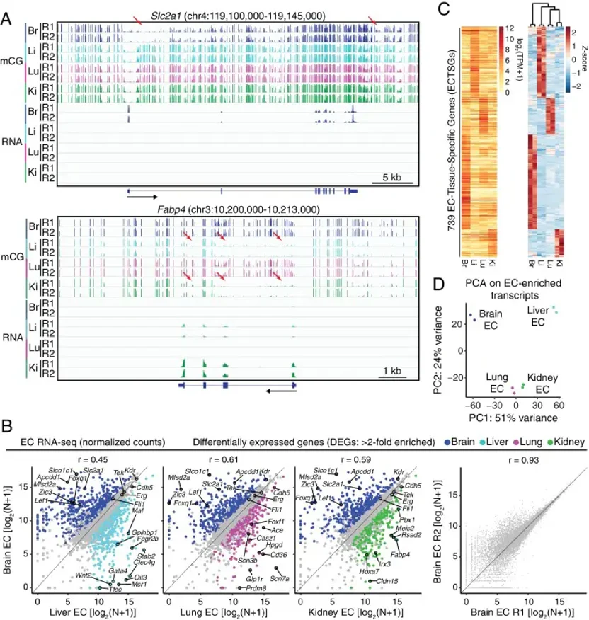
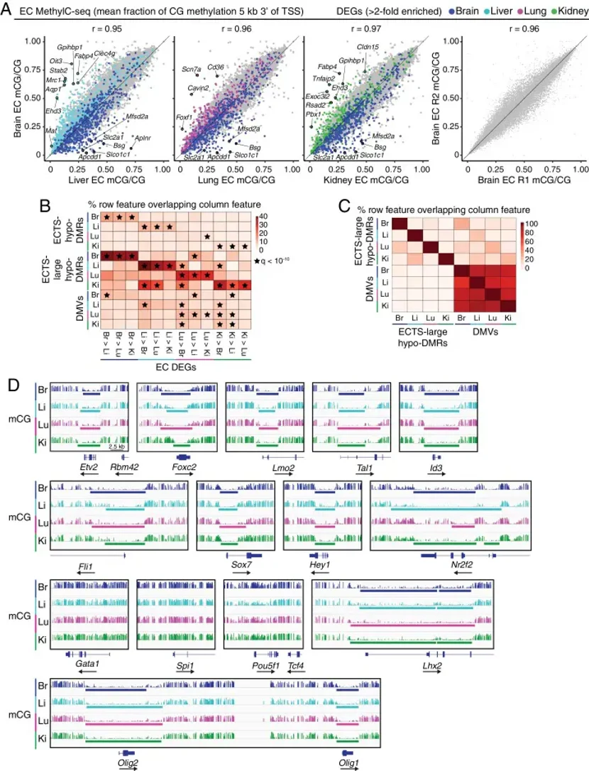
Organ-specific vascular endothelial cells (ECs) exhibit specialized functions, yet the underlying genomic mechanisms remain unclear. Sabbagh et al. generated a multi-omic atlas of mouse ECs isolated from brain, liver, lung, and kidney at postnatal day 7 (P7).
ECs were purified from Tie2-GFP mice via FACS (Figure 1). Although Tie2-GFP expression was absent in specific microvascular beds (e.g., renal glomeruli, Figure 1), the isolation protocol yielded high-purity populations suitable for multi-omic profiling. They performed RNA-seq, MethylC-seq, and ATAC-seq to map transcriptomes, DNA methylomes, and accessible chromatin landscapes across tissues. All experiments utilized multiple biological replicates, which exhibited high pairwise correlations (Figure 1 and 2).
Analysis of transcription factor (TF) expression and cis-regulatory motifs revealed both conserved and organ-specific EC regulatory networks. This comprehensive dataset, termed the Vascular Endothelial Cell Trans-omics Resource Database (VECTRDB), is publicly accessible for exploration.
Ask a Question
Write your own review
Description: C57BL/6-GFP Mouse Skeletal Muscle Microvascular Endothelial Cells from Creative Bioarray are isolated from C57BL/6-Tg (CAG-EGFP) 1Osb/J mouse skeletal muscle tissue of pathogen-free laboratory mice. ...
Description: eNOS KO Mouse Stomach Epithelial Cells from Creative Bioarray are isolated from stomach tissue of pathogen-free laboratory mice. eNOS KO Mouse Stomach Epithelial Cells are grown in a T25 tissue ...
Description: eNOS KO Mouse Liver Fibroblasts from Creative Bioarray are isolated from liver tissue of pathogen-free laboratory mice. eNOS KO Mouse Liver Fibroblasts are grown in T75 tissue culture flasks ...
Description: C57BL/6-GFP Mouse Corneal Epithelial Cells from Creative Bioarray are isolated from C57BL/6-GFP-Tg(CAG-EGFP)1Osb/J mouse corneal tissue of pathogen-free laboratory mice. C57BL/6-GFP Mouse Corneal ...

Description: BALB/c Mouse Retinal Microvascular Endothelial Cells from Creative Bioarray are isolated from retinal tissue of pathogen-free laboratory mice. BALB/c Mouse Retinal Microvascular Endothelial Cells are ...