
42-MG-BA
Cat.No.: CSC-C0466
Species: Homo sapiens (Human)
Source: Brain
Morphology: flat epithelial-like polygonal cells growing adherently as monolayer or, if confluent, as multilayers with processes
Culture Properties: monolayer
- Specification
- Background
- Scientific Data
- Q & A
- Customer Review
Immunology: cytokeratin -, desmin -, endothel -, GFAP +, neurofilament -, vimentin +
Viruses: ELISA: revers
The 42-MG-BA cell line is a continuous human glioblastoma (GBM) line established from the surgical specimen of a 67-year-old male patient with primary, treatment-naïve WHO grade IV glioma. Because the tumor was resected prior to any radio- or chemotherapy, the cell line preserves the native genomic landscape of primary GBM, free from therapy-induced artifacts. This makes it a particularly authentic model for dissecting de novo gliomagenesis and intrinsic therapeutic resistance.
A key molecular hallmark of 42-MG-BA is a homozygous missense mutation in TP53 at codon 273 (R273H), one of the most frequent gain-of-function hotspot mutations in human cancer. Importantly, the cells harbor wild-type IDH1/2 and lack EGFR amplification, defining a distinct molecular subset of classical GBM. They exhibit spindle-shaped, adherent morphology with robust expression of the glial markers GFAP and vimentin, confirming astrocytic lineage fidelity. When implanted orthotopically into immunodeficient mice, 42-MG-BA forms highly invasive, diffusely infiltrating tumors that faithfully recapitulate the histological hallmarks of human GBM, including perivascular pseudorosettes, microvascular proliferation, and zones of necrosis.
The stable phenotype and well-defined genetic background of 42-MG-BA ensure high experimental reproducibility. Combined with its ability to generate aggressive in vivo tumors, this line serves as an invaluable platform for studying p53 gain-of-function biology, mechanisms of diffuse brain invasion, tumor-stroma interactions, and for preclinical evaluation of anti-invasive and anti-glioma therapies.
Radiotherapy-Activated NBTXR3 Nanoparticles Induce Lysosomal Membrane Permeabilization
Radiotherapy-activated NBTXR3 (NBTXR3 + RT) has demonstrated superior efficacy in cancer cell destruction and tumor growth control, compared to radiotherapy (RT), in preclinical and clinical settings. However, the early events leading to these results, such as NBTXR3 endocytosis, intracellular trafficking and primary biological responses induced by NBTXR3 + RT remain poorly understood.
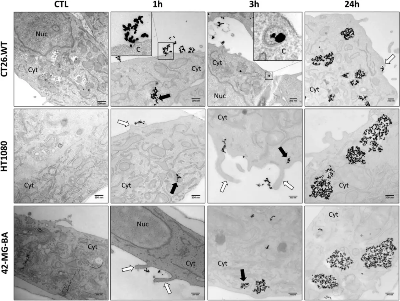
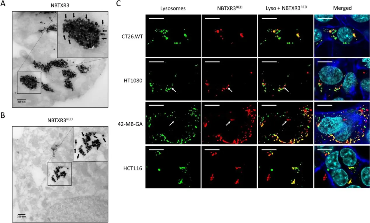
We analyzed by transmission electron microscopy endocytosis and intracellular localization of NBTXR3 nanoparticles after endocytosis in various cell lines, in vitro and in vivo. A kinetic of NBTXR3 endocytosis and its impact on lysosomes was conducted using LysoTracker staining, and a RNAseq analysis was performed.
NBTXR3 nanoparticles were rapidly internalized by cells mainly through macropinocytosis and in a less extend by clathrin-dependent endocytosis. NBTXR3-containing endosomes were then fused with lysosomes. The day following NBTXR3 addition, we measured a significant increase in LysoTracker lysosome labeling intensity, in vitro as in vivo. Following RT, a significant lysosomal membrane permeabilization (LMP) was measured exclusively in cells treated with NBTXR3 + RT, while RT had no effect.
Ask a Question
Write your own review
- You May Also Need
Description: Established from the biopsy specimens from a 42-year-old woman with glioma (histologically defined as glioblastoma)
Description: Established from the frontal brain lobe of a 54-year-old woman with glioblastoma multiforme in 1989 (pre-treatment)
Description: Derived from an adult female with glioblastoma multiforme treated with local brain irradiation and multi drug chemotherapy. By flow cytometry these cells were almost 100% positive for MHC Class I.
Description: 1321N1 is a human astrocytoma cell line isolated in 1972 as a sub clone of the cell line 1181N1 which in turn was isolated from the parent line U-118 MG (one of a number of cell lines derived from ...
- Adipose Tissue-Derived Stem Cells
- Human Neurons
- Mouse Probe
- Whole Chromosome Painting Probes
- Hepatic Cells
- Renal Cells
- In Vitro ADME Kits
- Tissue Microarray
- Tissue Blocks
- Tissue Sections
- FFPE Cell Pellet
- Probe
- Centromere Probes
- Telomere Probes
- Satellite Enumeration Probes
- Subtelomere Specific Probes
- Bacterial Probes
- ISH/FISH Probes
- Exosome Isolation Kit
- Human Adult Stem Cells
- Mouse Stem Cells
- iPSCs
- Mouse Embryonic Stem Cells
- iPSC Differentiation Kits
- Mesenchymal Stem Cells
- Immortalized Human Cells
- Immortalized Murine Cells
- Cell Immortalization Kit
- Adipose Cells
- Cardiac Cells
- Dermal Cells
- Epidermal Cells
- Peripheral Blood Mononuclear Cells
- Umbilical Cord Cells
- Monkey Primary Cells
- Mouse Primary Cells
- Breast Tumor Cells
- Colorectal Tumor Cells
- Esophageal Tumor Cells
- Lung Tumor Cells
- Leukemia/Lymphoma/Myeloma Cells
- Ovarian Tumor Cells
- Pancreatic Tumor Cells
- Mouse Tumor Cells