Porcine Lung Fibroblasts
Cat.No.: CSC-C4893L
Species: Pig
Source: Lung
Cell Type: Fibroblast
- Specification
- Background
- Scientific Data
- Q & A
- Customer Review
Never can cryopreserved cells be kept at -20 °C.
Porcine Lung Fibroblasts are primary cells derived from porcine lung parenchyma. They are frequently used as a lung model in vitro to study normal pulmonary structure, repair mechanisms, and disease pathophysiology. Fibroblasts are one of several major stromal cell types present within the lung parenchyma. In vivo, fibroblasts play a major role in regulating ECM homeostasis and contribute to tissue maintenance and injury responses.
The predominant role of fibroblasts in vivo is to synthesize and remodel ECM proteins including collagen, fibronectin, and elastin. Fibroblasts also engage in wound healing responses, and upon profibrotic stimulation, can differentiate into myofibroblasts. Porcine lung fibroblasts are especially useful to study because pigs have many similarities to human lung physiology. Under standard culture conditions, Porcine Lung Fibroblasts exhibit a spindle-shaped adherent morphology and will proliferate. These cells have been used to study pulmonary fibrosis, inflammatory signaling pathways, viral infections, toxicology, and tissue engineering applications. They have also been used as a supportive stromal cell layer for organotypic cultures and air-liquid interface cultures to study fibroblast-epithelial cell interactions.
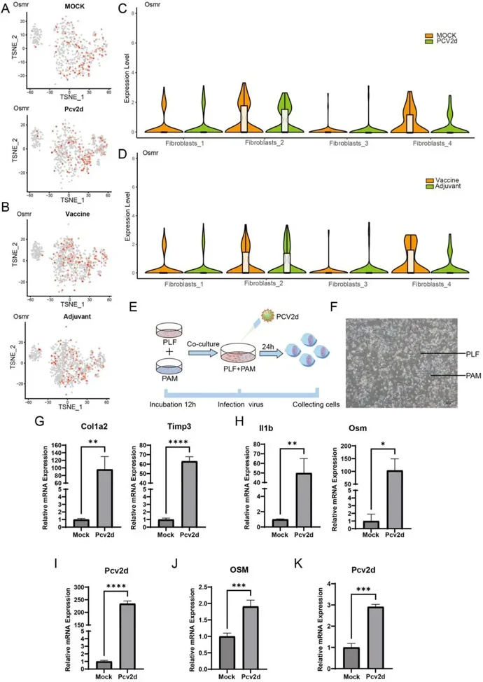
In Vitro Co-Culture Model Validates the Fibrotic Regulatory Mechanism
Pulmonary fibrosis involves aberrant lung fibroblast activation producing excessive extracellular matrix. Xu's team investigated how PCV2d viral infection induces fibroblast activation through alveolar macrophage-derived oncostatin M (OSM), aiming to identify therapeutic targets for viral-induced pulmonary fibrosis.
To verify pulmonary fibrosis regulatory mechanisms in a cross-species model, they constructed a porcine macrophage-lung fibroblast (PAM-PLF) heterologous co-culture system (Fig. 1E). Single-cell sequencing identified Osmr, the OSM receptor, on PLFs (Fig. 1A-D). Following PCV2d virus inoculation, viral load and fibrosis markers were assessed by qRT-PCR at 24 hours (Fig. 1G-I). PCV2d levels increased significantly post-infection, confirming successful model establishment. PCV2d infection significantly activated fibrosis-related molecular networks, with marked upregulation of COL1A2 and TIMP3 (Fig. 1G-H). Key inflammatory mediators IL1b and OSM also increased significantly. To identify the OSM source, PAMs and PLFs were cultured separately with PCV-2 virus. qRT-PCR at 24 hours showed OSM was predominantly expressed in PAMs, with negligible PLF expression (Fig. 1J-K), confirming macrophages as the primary OSM source in this context.
At present, cell freezing mostly uses DMSO dimethyl sulfoxide or glycerol as a protective agent, and there are many kinds of cell freezing solution formulations, such as medium: serum: DMSO = 7:2:1 or 8:1:1 or 5:4:1, or directly with serum: DMSO = 9:1, and generally a high concentration of serum helps to maintain cell viability, and the survival rate of resuscitation is more than 80% to 90%.
Ask a Question
Average Rating: 4.0 | 1 Scientist has reviewed this product
Consistent results
We were able to get consistent results from the cell products in our experiments.
12 Aug 2023
Ease of use
After sales services
Value for money
Write your own review
Description: Porcine Corneal Epithelial Cells from Creative Bioarray are isolated from corneal tissue of porcine. Porcine Corneal Epithelial Cells are grown in a T25 tissue culture flask pre-coated with ...
Description: Porcine Brain Vascular Fibroblasts from Creative Bioarray are isolated from brain tissue of porcine. Porcine Brain Vascular Fibroblasts are grown in T75 tissue culture flasks pre-coated with ...
Description: Porcine Thymus Endothelial Cells from Creative Bioarray are isolated from thymus tissue of porcine. Porcine Thymus Endothelial Cells are grown in T25 tissue culture flasks pre-coated with ...

Description: Porcine Kidney Endothelial Cells from Creative Bioarray are isolated from kidney tissue of porcine. Porcine Kidney Endothelial Cells are grown in T25 tissue culture flasks pre-coated with ...

Description: Pig bone marrow from Creative Bioarray is procured from the fresh femurs. Bone Marrow-CD34+ stem/progenitor cells are positively isolated using a direct immunomagnetic CD34 MicroBead labeling system.
Description: Porcine Primary Thymus Fibroblasts from Creative Bioarray are isolated from thymus tissue of porcine. Porcine Primary Thymus Fibroblasts are grown in T75 tissue culture flasks pre-coated with ...