
Human Uterine Epithelial Cells
Cat.No.: CSC-C4880L
Species: Human
Source: Uterus
Cell Type: Epithelial Cell
- Specification
- Background
- Scientific Data
- Q & A
- Customer Review
Never can cryopreserved cells be kept at -20 °C.
Human Uterine Epithelial Cells are primary cells isolated from the endometrial or cervical tissue of the human uterus. They compose the superficial lining of the uterine wall and are key players in reproductive physiology such as the menstrual cycle, embryonic implantation, and barrier defence against pathogens. Morphologically, these cells show a classical epithelial phenotype, generally presenting as a monolayer of adherent polygonal or cobblestone-shaped cells.
In vitro, they are cultured in a nutrient-rich basal medium supplemented with serum and essential amino acids to support proliferation and maintain phenotypic stability. Standard culture conditions require a humidified incubator at 37°C with 5% carbon dioxide. Normal protease solution is normally used for confluence with subculture.
Human Uterine Epithelial Cells are physiologically relevant and serve as an important in vitro model for the study of endometrial biology, hormone responses (e.g., to oestrogen and progesterone), maternal-fetal interactions and the pathogenesis of uterine disorders such as endometriosis, fibrosis and gynaecological cancers. In addition, they are widely used to evaluate drug efficacy and toxicity in the female reproductive tract.
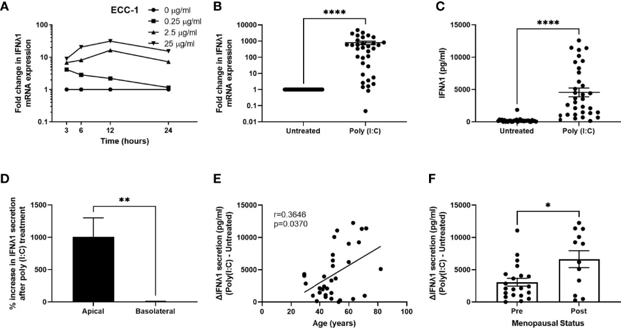
Poly (I:C) Induces IFNλ1 Expression and Secretion by Epithelial Cells
It is well known that estradiol (E2) and progesterone (P) modulate immune function in the uterine endometrium but their role in regulating IFNλ1 expression in the non-pregnant uterus has not been understood. To study this Patel et al. used purified cultures of human uterine epithelial and stromal cells.
While poly (I:C) has been shown to induce a strong innate immune response in female reproductive tract (FRT) cells previously, the specific function of IFNλ1 has not been elucidated. They found that poly (I:C) induced IFNλ1 mRNA in a dose-dependent manner with a maximum response at 12-24 h after stimulation with 25 µg/ml (Fig. 1A) in polarised ECC-1 epithelial cells. Poly (I:C) treatment of primary uterine epithelial cells for 24 hours induced a strong response, with IFNλ1 mRNA up-regulation averaging 790-fold (Fig. 1B) and apical secretion increasing to about 4500 pg/ml (Fig. 1C). Basolateral IFNλ1 levels were not altered (Fig. 1D), and apical constitutive secretion was relatively low (~20.5 pg/ml, Fig. 1D). There was considerable inter-patient variability with mRNA fold-changes from 0.04 to 4800 and apical secretion of 120-12300 pg/ml.
After the cell culture medium is prepared, a small amount should be extracted and put into the culture bottle and placed in the incubator at 37℃ for 24-48 h to check whether the culture medium is contaminated or not, then it can be used for experiments. The serum required for the preparation of culture medium should be qualified and kept stable. After a batch of good results, you can buy more serum of the same batch number, so that the experimental conditions are stable, and it is easier to adjust the pH value of the liquid.
Ask a Question
Average Rating: 5.0 | 1 Scientist has reviewed this product
Multiple purchases
After purchasing the LA-N-1, I have also purchased other products and services from Creative Bioarray several times.
05 Apr 2022
Ease of use
After sales services
Value for money
Write your own review
Description: GFP Expressing Human Uterine Microvascular Endothelial Cells (GFP-HUMECs) provided by Creative Bioarray are puromycin-selected from Human Uterine Microvascular Endothelial Cells infected with ...
Description: Human Primary Sertoli Cells are isolated form normal human prostate tissue. T25 flasks is required for cell adhension to the culture vessels. Grow cells in ECM-coated culture vessels with 5% CO2. ...

Description: Human Testicular Endothelial Cells are isolated form normal human tesis tissue. T25 flasks is required for cell adhension to the culture vessels. Grow cells in ECM-coated culture vessels with 5% CO2. ...
Description: Origin: HumanDesease: Robertsonian Translocation (RTL)Application: These cells can be used to investigate reproduction, stem cells, disease in vitro and developmental studies.
Description: Human Ovarian Epithelial Cells are isolated from normal human ovary tissue.