
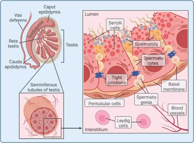
Human Sertoli Cells
Cat.No.: CSC-C9380W
Species: Human
Source: Testis
Morphology: Bipolar
Cell Type: Sertoli Cell
- Specification
- Background
- Scientific Data
- Q & A
- Customer Review
Human Sertoli Cells are a primary somatic cell line derived from human testicular tissue and utilized as an in vitro model that more closely represents physiological responses of the testis and allows for study of male reproductive biology. Sertoli Cells are somatic cells found within the seminiferous tubules of the testis that support spermatogenesis.
Under typical culture conditions, human Sertoli cells appear elongated or irregular polygonal and are adherent, growing as a monolayer. Cells display positive staining for known Sertoli cell markers such as SOX9, GATA4, WT1, vimentin, and androgen receptor (AR) demonstrating cell lineage. Sertoli Cells form tight junction complexes and have been shown to respond to hormones such as follicle-stimulating hormone (FSH) and testosterone. Human Sertoli Cells have been used as a model system to study regulation of spermatogenesis as well as development of the testis and pathogenesis of male infertility. Researchers have employed human Sertoli Cells to explore the mechanisms of testicular toxicology and endocrine disruption as well as how testicular immune privilege functions. These cells have also been used in co-culture studies to better understand Sertoli-germ cell interactions or assess the impact of pharmaceuticals/environmental toxicants on spermatogenesis.
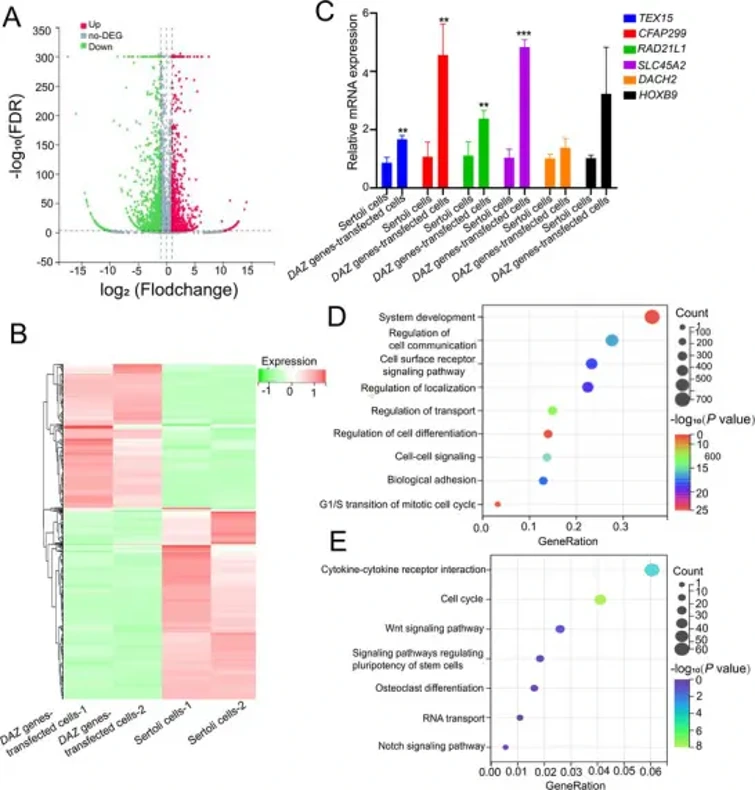
RAD21L1 Transcript is Upregulated by Overexpressing DAZ Family Genes during Reprogramming of Sertoli Cells into Human SSCs
Previously reported that primary human Sertoli cells can be reprogrammed into human spermatogonial stem cells (SSCs) phenotypically by overexpressing DAZ family genes (DAZ, DAZL, and BOULE). However, the molecular mechanisms underlying this transition remain unclear.
To identify key regulators, He et al. performed RNA-seq and found 1,972 differentially expressed genes (DEGs) between DAZ-transfected cells and control Sertoli cells. Volcano plots and heat maps revealed 911 upregulated and 1,061 downregulated genes (Fig. 1A, B). qPCR verified several upregulated candidates, including RAD21L1 (Fold change: 8.856, p = 0.0089), TEX15, CFAP299 (c40rf22), SLC45A2, DACH2, and HOXB9 (Fig. 1C). KEGG and GO analyses showed these DEGs were enriched in pathways regulating stem cell pluripotency, cell communication, and differentiation (Fig. 1D, E). Collectively, their data demonstrate that RAD21L1 is upregulated during DAZ-mediated reprogramming of Sertoli cells to human SSCs.
Nowadays, most laboratories will remove DMSO by adding culture medium after thawing, but some researchers believe that, except for a small number of cells specifically stated to be sensitive to DMSO, most cell lines (including suspension cells) can be thawed and placed into culture corner flasks containing 10-15 mL of fresh medium, and then replaced with fresh medium the next day to remove the DMSO, which can avoid the problem of most cells failing to grow or attach after thawing. This can avoid most of the problems that cells cannot grow or attach after thawing.
Ask a Question
Average Rating: 5.0 | 1 Scientist has reviewed this product
Good growth
The cells are in good growth condition when observed through a microscope.
14 July 2022
Ease of use
After sales services
Value for money
Write your own review

Description: GFP Expressing Human Uterine Microvascular Endothelial Cells (GFP-HUMECs) provided by Creative Bioarray are puromycin-selected from Human Uterine Microvascular Endothelial Cells infected with ...
Description: Human Uterine Epithelial Cells are isolated from normal human uterine tissue.
Description: Human Testicular Endothelial Cells are isolated form normal human tesis tissue. T25 flasks is required for cell adhension to the culture vessels. Grow cells in ECM-coated culture vessels with 5% CO2. ...
Description: Origin: HumanDesease: Robertsonian Translocation (RTL)Application: These cells can be used to investigate reproduction, stem cells, disease in vitro and developmental studies.
Description: Human Ovarian Epithelial Cells are isolated from normal human ovary tissue.