
Human Ovarian Epithelial Cells
Cat.No.: CSC-C4876L
Species: Human
Source: Ovary
Cell Type: Epithelial Cell
- Specification
- Background
- Scientific Data
- Q & A
- Customer Review
Never can cryopreserved cells be kept at -20 °C.
Human Ovarian Epithelial Cells (HOECs), also known as ovarian surface epithelial cells (OSE), are an important cell type derived from mesothelial tissue, closely associated with ovulation and serving as the primary cell of origin for the majority of ovarian cancers, particularly high-grade serous carcinoma. Recent studies have shown that normal and malignant ovarian epithelial cells differ in their response to growth factor stimulation, notably, an escape from inhibition by transforming growth factor-β. Studies on the biological properties of ovarian surface epithelial cells will be of great significance for understanding their transformation process.
HOECs are extensively used to investigate early genetic alterations (e.g., TP53 mutations), signaling pathways (like Wnt, Notch), tumor microenvironment interactions, and for developing novel therapeutic strategies, including targeted therapies and immunotherapies. The main advantage of using HOECs, especially primary cultures directly derived from patient tissue (normal, benign, or malignant), is their superior physiological relevance – they retain the patient's native genetic background, epigenetic landscape, and phenotypic heterogeneity, providing more accurate insights into human disease mechanisms and treatment responses compared to immortalized cell lines that often undergo genetic drift or adaptive modification. This makes them crucial for translational research aimed at improving ovarian cancer outcomes.
Human Ovarian Surface Epithelium Organoids as a Platform to Study Tissue Regeneration
Although the regenerative properties of the ovarian surface epithelium (OSE) have been well studied in mice, understanding the precise mechanism of tissue repair in the human ovary remains hampered by limited access to human ovaries and suitable in vitro culture protocols. Tissue-specific organoids, miniaturized in vitro models replicating both structural and functional aspects of the original organ, offer new opportunities for studying organ physiology, disease modeling, and drug testing.
This protocol describes establishing three-dimensional (3D) tissue organoids from primary human ovarian surface epithelium (hOSE) cells. The hOSE organoids exhibited (donor-specific) variability in morphology and growth, highlighting their utility for personalized studies. Additionally, this protocol includes hOSE organoid maintenance, passaging, and immunofluorescence within the same culture plate. Furthermore, it provides a description of the different morphology that hOSE organoids can adopt and characterizes changes in immunophenotype during culture. This method may enable the discovery of factors contributing to OSE regeneration and facilitate patient-specific drug screenings for malignant OSE.
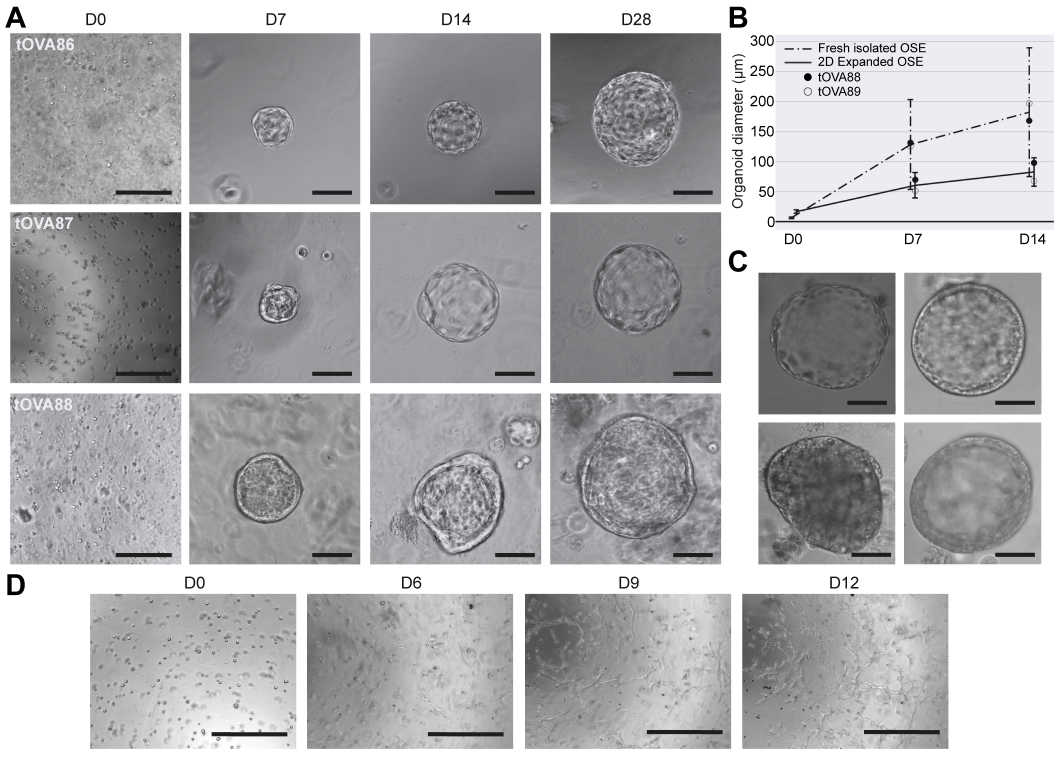 Fig. 1. Morphological characterization of hOSE organoids (Del Valle, Julieta S., et al. 2024).
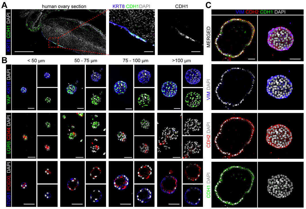 Fig. 2. Cellular characterization of hOSE organoids (Del Valle, Julieta S., et al. 2024).
Phenol red is used in culture media as an indicator of pH: red when neutral, yellow when acidic, and purple when basic. Studies have shown that phenol red mimics the effects of steroid hormones (especially estrogen). To avoid steroid reactions, culture cells, especially mammalian cells, in medium without phenol red. Because phenol red interferes with the assay, some researchers do not use media with phenol red when doing flow cytometry.
Ask a Question
Average Rating: 5.0 | 1 Scientist has reviewed this product
Reliable products
We have always relied on Creative Bioarray's products because they have helped us to complete our experiments well.
17 July 2022
Ease of use
After sales services
Value for money
Write your own review

Description: GFP Expressing Human Uterine Microvascular Endothelial Cells (GFP-HUMECs) provided by Creative Bioarray are puromycin-selected from Human Uterine Microvascular Endothelial Cells infected with ...

Description: Human Primary Sertoli Cells are isolated form normal human prostate tissue. T25 flasks is required for cell adhension to the culture vessels. Grow cells in ECM-coated culture vessels with 5% CO2. ...

Description: Human Uterine Epithelial Cells are isolated from normal human uterine tissue.
Description: Human Testicular Endothelial Cells are isolated form normal human tesis tissue. T25 flasks is required for cell adhension to the culture vessels. Grow cells in ECM-coated culture vessels with 5% CO2. ...
Description: Origin: HumanDesease: Robertsonian Translocation (RTL)Application: These cells can be used to investigate reproduction, stem cells, disease in vitro and developmental studies.