
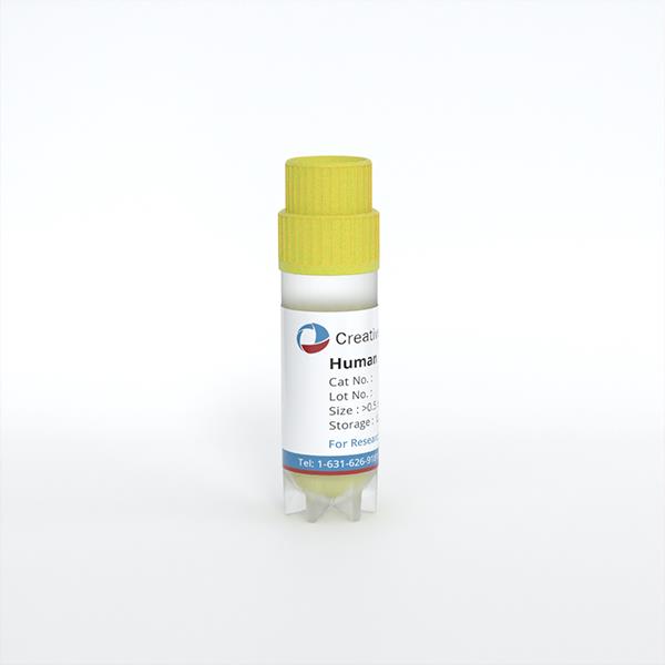
Human Liver Sinusoidal Microvascular Endothelial Cells
Cat.No.: CSC-C4863L
Species: Human
Source: Liver
Cell Type: Endothelial Cell; Microvascular Cell
- Specification
- Background
- Scientific Data
- Q & A
- Customer Review
Never can cryopreserved cells be kept at -20 °C.
Human liver sinusoidal microvascular endothelial cells (LSECs) are a specialized endothelial population lining the hepatic sinusoids, distinguished by the presence of transcellular fenestrations, the absence of a organized basement membrane, and a unique repertoire of scavenger receptors (e.g., Stabilin‑1/-2, LRP‑1, mannose receptor). These fenestrated, sieve‑like structures enable bidirectional passage of solutes and macromolecules between sinusoidal blood and hepatocytes, making LSECs central to liver metabolic regulation, lipid trafficking, and waste clearance.
Compared to vascular endothelial cells from large vessels (e.g., HUVEC), primary human LSECs offer several unique advantages for research and pharmaceutical applications:
- High endocytic clearance capacity - LSECs are the body's most efficient scavenger cells, contributing to >70 % of total liver clearance of soluble macromolecules, waste products (e.g., hyaluronic acid, AGEs), and modified plasma proteins. This property is essential for drug delivery studies and evaluation of nanoparticle‑based therapies.
- Immune‑privileged and tolerogenic functions - LSECs express PD‑L1, scavenging receptors, and cross‑present MHC I, inducing regulatory T cells and peripheral tolerance. They are therefore a powerful tool for modeling hepatic immune regulation and chronic inflammation.
- Direct involvement in fibrosis and portal hypertension - Upon activation, LSECs undergo capillarization, a critical step in fibrogenesis. Primary LSECs allow exact mechanistic studies of sinusoidal remodeling and drug screening for anti‑fibrotic strategies.
- Physiologically relevant drug metabolism and toxicity models - Co‑cultured with hepatocytes, LSECs recreate the native liver microenvironment, improving prediction of drug‑induced liver injury (DILI) and metabolism.
Vitamin D Attenuates Hepatic Sinusoidal Capillarization via Dual Autophagy Activation and Pyroptosis Suppression in Liver Sinusoidal Endothelial Cells
Metabolic dysfunction-associated fatty liver disease (MAFLD) is closely associated with type 2 diabetes mellitus (T2DM), where T2DM serves as a crucial driving factor for MAFLD progression. While vitamin D (VD) demonstrates protective effects against MAFLD, the underlying mechanisms through which it influences MAFLD-related liver sinusoidal endothelial cell (LSEC) capillarization remain to be elucidated. This study aimed to explore how vitamin D ameliorates LSEC capillarization in T2DM-associated MAFLD.
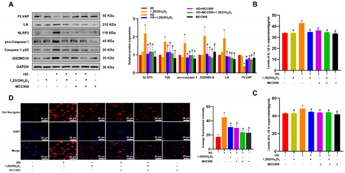
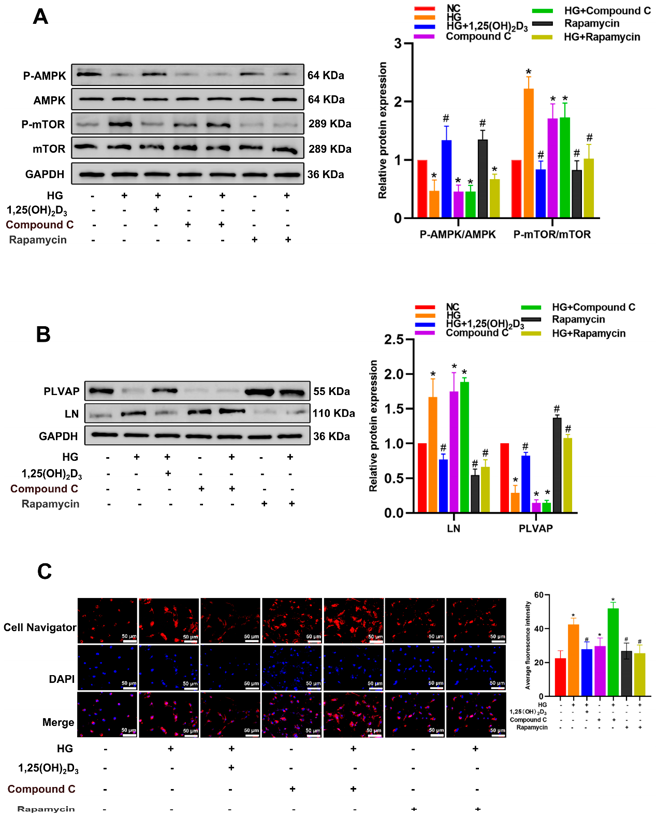
Culture human liver sinusoidal endothelial cells (HLSECs) according to the established protocol. After 1,25(OH)2D3 intervention in high glucose (HG)-induced HLSECs, determine the changes in liver sinusoidal capillarization-related proteins (LN, PLVAP), autophagy and pyroptosis levels. Observe the changes in cell lipid accumulation and fenestration structures. After adding Bafilomycin A1, MCC950, compound C and rapamycin to HLSECs, explore the therapeutic mechanism of 1,25(OH)2D3.
HG can induce the capillarization and lipid accumulation of HLSEC, increase the level of pyroptosis, and simultaneously reduce the autophagy level. Vitamin D alleviated high-glucose-induced pyroptosis (by suppressing GSDMD/NLRP3) and autophagic inhibition by activating the AMPK-mTOR axis (upregulating p-AMPK and downregulating mTOR), and improved lipid accumulation and hepatic sinusoidal capillarization. These results demonstrate, for the first time, that VD mitigates LSEC dysfunction through dual mechanisms: activating AMPK-dependent autophagy and inhibiting pyroptosis, providing a therapeutic rationale for VD in treating MAFLD-related sinusoidal pathology.
Ask a Question
Write your own review
Description: QualiCell® Human Kupffer Cells are isolated from adult human livers. Each vial contains a minimum of one million viable cells. These easy-to-use cells provide a convenient way to produce hepatocyte ...
Description: GFP Expressing Human Hepatic Sinusoidal Microvascular Endothelial Cells (GFP-HLiSMVECs) are puromycin-selected from Human Hepatic Sinusoidal Microvascular Endothelial Cells infected with lentivirus ...
Description: First, fetal liver is homogenized. Then, a density separation gradient media (1.077g/ml density) is used to isolate the Mononuclear Cells. Next, a direct immunomagnetic CD34 MicroBead labeling system ...
Description: The liver fulfills many vital processes in mammals. It is the central organ of energy metabolism, biotransformation of xenobiotics, and synthesis of plasma proteins under physiological and ...
Description: Human Liver Epithelial Cells are isolated from normal human liver tissue.
Description: Recent research indicates cancer associated fibroblasts (CAFs) significant involvement in crucial aspects of epithelial solid tumor biology, specifically neoplastic progression, tumor growth, ...