
Human Liver Cancer Associated Fibroblasts
Cat.No.: CSC-C30036J
Species: Human
Source: Liver
Cell Type: Fibroblast
- Specification
- Background
- Scientific Data
- Q & A
- Customer Review
Human Liver Cancer-Associated Fibroblasts (hCAFs) are primary stromal cells isolated directly from human hepatocellular carcinoma (HCC) specimens, representing the most abundant and functionally critical non-malignant constituents of the tumor microenvironment (TME). Unlike immortalized cell lines or animal xenografts, hCAFs preserve patient-specific genetic and epigenetic landscapes, sustain native activated myofibroblast phenotypes, and authentically recapitulate the intricate paracrine signaling networks that govern HCC progression, desmoplasia, and therapy resistance.
Predominantly derived from activated hepatic stellate cells, hCAFs constitutively express established CAF markers—including FAP-α, α-SMA, vimentin, and PDGFR-β—while secreting a potent array of tumor-modulating mediators such as TGF-β1, IL-6, HGF, and SDF-1/CXCL12. Through dynamic extracellular matrix remodeling and bidirectional crosstalk with neoplastic and immune compartments, they actively promote cancer cell proliferation, angiogenesis, epithelial-mesenchymal transition, and the establishment of an immunosuppressive niche, faithfully mirroring the pathophysiological complexity of the human HCC microenvironment in vivo. Contemporary single-cell transcriptomic and spatial omics analyses have further unveiled substantial inter-patient heterogeneity among hCAF subpopulations, highlighting their indispensable value in personalized biomarker discovery, patient stratification, and subtype-specific therapeutic targeting.
As a physiologically relevant ex vivo platform, hCAFs offer distinct and unparalleled experimental advantages: native secretory fidelity, superior translational predictability across diverse in vitro systems, and seamless compatibility with co-culture platforms, 3D organoid models, and high-throughput drug screening workflows. Moreover, their relative genetic stability compared to malignant cells renders them robust, druggable targets for mechanism-driven validation and stroma-directed therapeutic discovery. Collectively, these attributes position hCAFs as an essential and versatile resource for advancing precision oncology, accelerating drug development, and informing personalized HCC treatment strategies.
CAFs Promote CSC Stemness Capacity via Extracellular Vesicles in Hepatocellular Carcinoma (HCC)
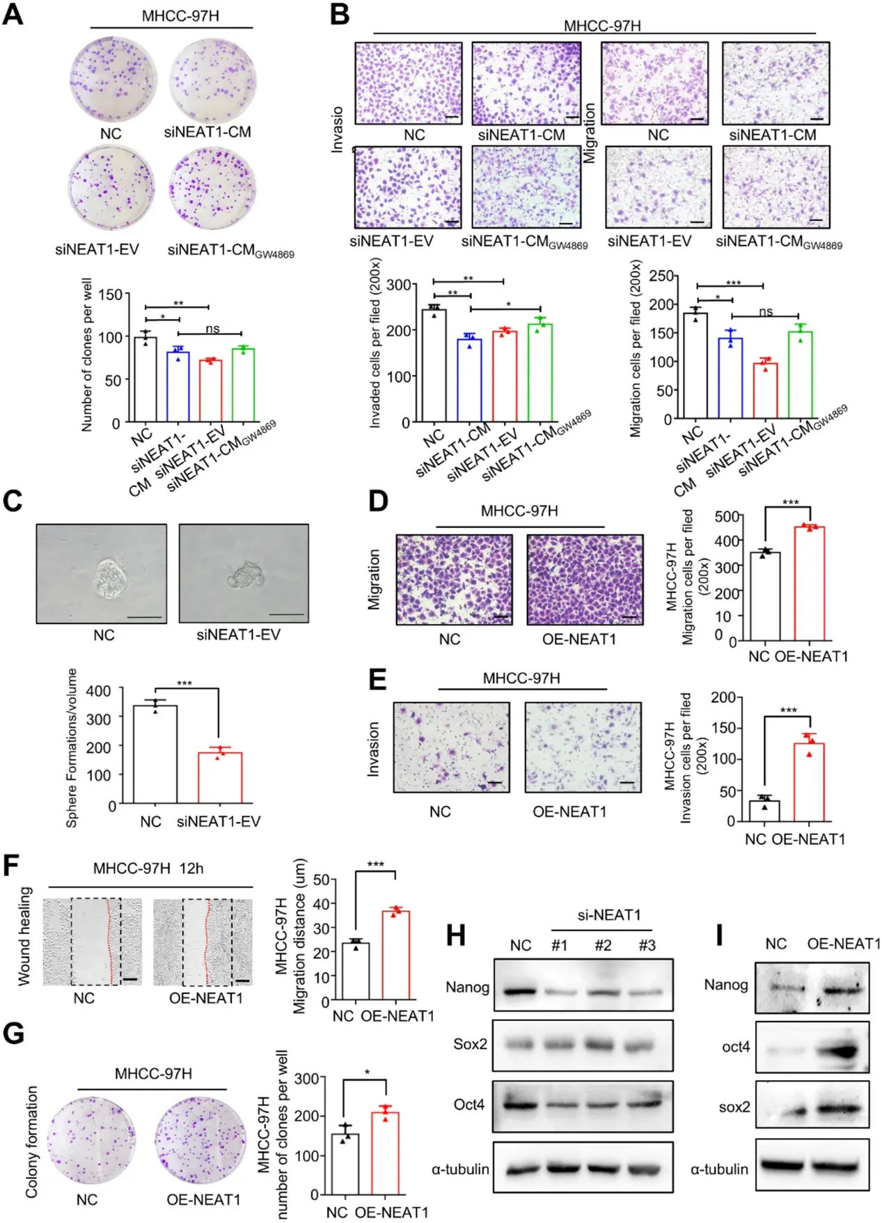
In this study, Chen, Wei, et al. identified a novel long non-coding RNA (lncRNA) NEAT1 in cancer-associated fibroblast (CAFs)-derived extracellular vesicles (EVs) that play a critical role in the induction of CSCs and HCC tumorigenesis.
To further elucidate the role of NEAT1 in cell stemness capacity, they employed siRNAs to down-regulate NEAT1 in CAFs. The cell culture supernatant (siNEAT1-CM), EVs (siNEAT1-EV), and CAFs conditioned medium treated with EVs release inhibitor GW4869 (siNEAT1-CM GW4869) were applied to MHCC-97 H and SNU398 cells. Transwell migration and cloning formation assays revealed a decrease in the migration of both cells in the siNEAT1-EV group and siNEAT1-CM group, which was significantly rescued when HCC cells were treated with GW4869 (Fig. 1A and B). Similarly, 3D spheroid formation assays indicated that siNEAT1 decreased the stemness capacity of HCC cells (Fig. 1C).
Conversely, they overexpressed NEAT1 in MHCC-97 H and SNU-398 cells by transfecting NEAT1 plasmids. Transwell migration/invasion and wound-healing assays demonstrated that NEAT1 overexpression significantly increased the migration and invasion of both HCC cell lines (Fig. 1D and F). Colony formation assays further indicated that NEAT1 overexpression enhanced cell growth and anchorage-independent growth of HCC cells (Fig. 1G). Western blot and qRT-PCR demonstrated that knockdown of NEAT1 decreased the expression levels of the cancer stemness-associated genes Nanog, Sox2, and Oct4 (Fig. 1H and I). Collectively, these findings suggest that NEAT1 plays a critical role in cell motility and stemness capacity.
Ask a Question
Write your own review
Description: The liver fulfills many vital processes in mammals. It is the central organ of energy metabolism, biotransformation of xenobiotics, and synthesis of plasma proteins under physiological and ...
Description: Fetal liver is dissociated in an asceptic manner, followed by density separation to enrich the mononuclear cells. The enriched cells are frozen using our optimized cryopreservation medium. Fetal ...
Description: Fetal liver is dissociated in an asceptic manner, followed by density separation to enrich the mononuclear cells. The enriched cells are frozen using our optimized cryopreservation medium. Fetal ...
Description: Human Liver Carcinoma Epithelial Cells from Creative Bioarray are isolated from human liver tumor tissue. Human Liver Carcinoma Epithelial Cells are grown in T25 tissue culture flasks pre-coated with ...
Description: Endothelial cells play multiple physiological functions and are central to many pathological processes. The liver contains two distinct endothelial cell types: vascular and sinusoidal. Sinusoidal ...
Description: Creative Bioarray's Pooled Human Liver Microsomes provide a fully characterized, relevant, reliable and reproducible in vitro tool. Pooled of 50 donors give an economic way of deriving an accurate ...