Hamster Small Intestinal Smooth Muscle Cells
Cat.No.: CSC-C4769L
Species: Hamster
Source: Small Intestine; Intestine
Cell Type: Smooth Muscle Cell
- Specification
- Background
- Scientific Data
- Q & A
- Customer Review
Never can cryopreserved cells be kept at -52 °C.
Hamster Small Intestinal Smooth Muscle Cells are main smooth muscle cells derived from the small intestine of healthy hamsters. These cells constitute one of the principal contractile cell populations of the intestinal wall and are necessary for the regulation of gastrointestinal motility, luminal transport and coordinated peristaltic activity. In vivo, intestinal smooth muscle cells are organized into circular and longitudinal muscular layers that operate in concert to control movement and mixing of the intestinal contents.
Cultured hamster small intestine smooth muscle cells proliferate as adherent cells with an elongated spindle-shaped appearance. They display classic smooth muscle markers, such as α-smooth muscle actin (α-SMA), smooth muscle myosin heavy chain (SM-MHC) and calponin, suggesting their contractile phenotype. Like smooth muscle cells of other gastrointestinal tissues, these cells respond to neurotransmitters, growth hormones, inflammatory mediators and mechanical stimuli that regulate cellular contraction, proliferation and phenotypic modification.
These cells are a good in vitro model for the study of processes of gastrointestinal motility, smooth muscle physiology, calcium signaling and cell-matrix interactions. They may also be used in studies of intestinal remodeling, inflammatory reactions, smooth muscle differentiation and pharmacological evaluation of drugs affecting gastrointestinal function. Hamsters are often used in investigations of infectious disease and gastrointestinal biology, therefore these cells may also prove useful for comparative studies of intestinal biology and host tissue responses under controlled experimental conditions.
Characterization of Hamster Bone Marrow-Derived Mesenchymal Stem Cells for Therapeutic Use
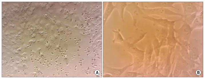
Bone marrow-derived mesenchymal stem cells (BM-MSCs) possess multilineage differentiation potential and secrete anti-inflammatory and trophic factors, making them promising candidates for cell therapy. In this study, BM-MSCs isolated from hamsters exhibited a typical fibroblast-like, spindle-shaped morphology upon attachment and reached 80% confluence within 3-4 days of incubation (Fig. 1).
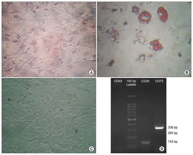
Functional assays confirmed their trilineage differentiation capacity. Under osteogenic induction, cells stained positive for Alizarin Red, verifying mineralized matrix formation (Fig. 2A). Adipogenic differentiation yielded intracellular lipid droplets, visualized by Oil Red O staining (Fig. 2B). Control cultures maintained in standard growth medium retained their fibroblast-like morphology and lacked evidence of osteogenic or adipogenic differentiation (Fig. 2C).
Immunophenotypic analysis by RT-PCR confirmed the mesenchymal identity of passage 3 BM-MSCs, with positive expression of CD29 and CD73 (mesenchymal markers) and absence of CD45 (hematopoietic marker) (Fig. 2D). These findings verify the successful isolation and characterization of functionally competent BM-MSCs, supporting their application in evaluating therapeutic strategies for busulfan-induced azoospermia.
Ask a Question
Write your own review
- You May Also Need
Description: Hamster Ovarian Smooth Muscle Cells are isolated from ovarian tissue of pathogen-free laboratory mice.
Description: Hamster Esophageal Epithelial Cells from Creative Bioarray are isolated from esophageal tissue of pathogen-free laboratory mice. Hamster Esophageal Epithelial Cells are grown in a T25 tissue culture ...
Description: Hamster Spleen Epithelial Cells from Creative Bioarray are isolated from spleen tissue of pathogen-free laboratory mice. Hamster Spleen Epithelial Cells are grown in a T25 tissue culture flask ...
Description: Hamster Corneal Epithelial Cells from Creative Bioarray are isolated from corneal tissue of pathogen-free laboratory mice. Hamster Corneal Epithelial Cells are grown in a T25 tissue culture flask ...
Description: Hamster Aortic Smooth Muscle Cells are isolated from aorta of hamster.
Description: Hamster Primary Pancreatic Fibroblasts are isolated from pancreas tissue of hamster.