
STC-1
Cat.No.: CSC-C9250W
Species: Mus musculus (Mouse)
Source: Intestine; Small Intestine; Duodenum
Morphology: epithelial-like
- Specification
- Background
- Scientific Data
- Q & A
- Customer Review
STC-1 is a murine intestinal enteroendocrine cell line originally established in 1990 from an endocrine tumor of the small intestine in double-transgenic mice harboring rat insulin promoter-driven polyoma small T antigen and SV40 large T antigen transgenes. As one of the three most extensively characterized enteroendocrine cell models, STC-1 offers a uniquely plurihormonal secretory repertoire, producing and releasing cholecystokinin (CCK), secretin, glucose-dependent insulinotropic polypeptide (GIP), peptide YY (PYY), neurotensin, and proglucagon-derived peptides including GLP-1, GLP-2, and oxyntomodulin in a regulated, stimulus-dependent manner.
This broad hormonal profile closely mirrors the overlapping hormone expression patterns of native upper gastrointestinal enteroendocrine cells, making STC-1 exceptionally relevant for investigating gut-brain signaling, nutrient sensing, and metabolic regulation. The cells mount robust, dose-dependent secretory responses to diverse physiological stimuli—including monosaccharides, fatty acids, amino acids, and bioactive peptides—providing a reliable platform for high-throughput screening of food-derived bioactives, satiety modulators, and incretin secretagogues. With decades of peer-reviewed validation and standardized culture protocols, STC-1 remains the gold-standard murine model for mechanistic studies of intestinal hormone secretion and therapeutic discovery in metabolic disease.
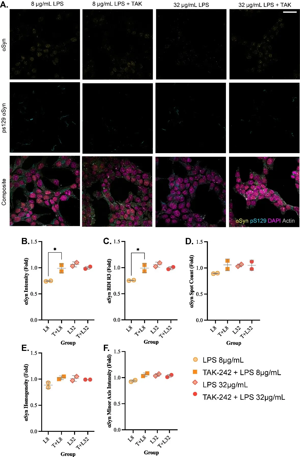
LPS Significantly Alters α-synuclein Intensity in A TLR4-Dependent Manner in STC-1 Cells
The STC-1 cell line, which models an enteroendocrine population capable of communicating with the gut microbiota, immune and nervous systems, was treated with a TLR4 inhibitor (TAK-242) prior to microbial lipopolysaccharide (LPS) exposure to investigate the role of TLR4 signaling in α-synuclein alterations. Antibodies targeting the full-length protein (α-synuclein) and the Serine-129 phosphorylated form (pS129) were used.
Confocal microscopy revealed heterogenous distribution of α-synuclein and pS129 in STC-1 cells, with prominent pS129 staining along cytoplasmic processes. Imaging flow cytometry further quantified the relationship between various α-synuclein morphometric features. Thereafter, imaging flow cytometry demonstrated a dose-specific effect of LPS, where the low (8 μg/mL), but not high dose (32 μg/mL), significantly altered measures related to α-synuclein intensity, distribution, and localization. Pre-treatment with a TLR4 inhibitor TAK-242 alleviated some of these significant alterations. This study demonstrates that LPS-TLR4 signaling alters the intracellular localization of α-synuclein in enteroendocrine cells.
Ask a Question
Write your own review
- You May Also Need
Description: neuroglial and neuronal character coexpressing ependymoma cell line.
Description: Established by irradiation of the adherent cells in long-term bone marrow cultures derived from C3H/HeNSlc strain mice
Description: Cloned from the C1300 mouse neuroblastoma line. Adapted to culture by alternate in vivo and in vitro passage. Cells have characteristics of mature neurons and secrete the enzymes for the synthesis of ...
Description: FL83B is a spontaneously immortalized hepatocyte cell line derived from the liver of a 15 to 17-day-old fetal C57BL/6J mouse.
Description: EMT6 was established from a transplantable murine mammary carcinoma that arose in a BALB/cCRGL mouse after implantation of a hyperplastic mammary alveolar nodule.
- Adipose Tissue-Derived Stem Cells
- Human Neurons
- Mouse Probe
- Whole Chromosome Painting Probes
- Hepatic Cells
- Renal Cells
- In Vitro ADME Kits
- Tissue Microarray
- Tissue Blocks
- Tissue Sections
- FFPE Cell Pellet
- Probe
- Centromere Probes
- Telomere Probes
- Satellite Enumeration Probes
- Subtelomere Specific Probes
- Bacterial Probes
- ISH/FISH Probes
- Exosome Isolation Kit
- Human Adult Stem Cells
- Mouse Stem Cells
- iPSCs
- Mouse Embryonic Stem Cells
- iPSC Differentiation Kits
- Mesenchymal Stem Cells
- Immortalized Human Cells
- Immortalized Murine Cells
- Cell Immortalization Kit
- Adipose Cells
- Cardiac Cells
- Dermal Cells
- Epidermal Cells
- Peripheral Blood Mononuclear Cells
- Umbilical Cord Cells
- Monkey Primary Cells
- Mouse Primary Cells
- Breast Tumor Cells
- Colorectal Tumor Cells
- Esophageal Tumor Cells
- Lung Tumor Cells
- Leukemia/Lymphoma/Myeloma Cells
- Ovarian Tumor Cells
- Pancreatic Tumor Cells
- Mouse Tumor Cells