
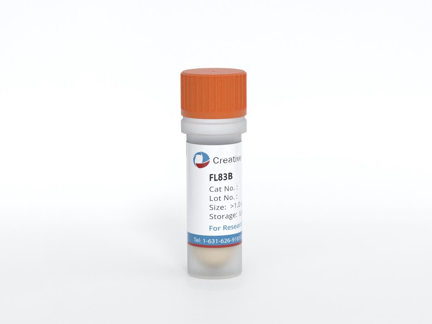
FL83B
Cat.No.: CSC-C7104J
Species: Mus musculus (Mouse)
Source: Embryo; Liver
Morphology: Epithelial
Culture Properties: Adherent
- Specification
- Background
- Scientific Data
- Q & A
- Customer Review
FL83B is an established murine hepatocyte cell line derived from normal fetal mouse liver at 15–17 days of gestation. The cells exhibit epithelial morphology and adherent growth, providing an expandable model for investigating selected aspects of hepatic biology. Although originating from normal tissue, FL83B cells are aneuploid and should be characterized as an established cell line rather than normal primary hepatocytes.
A distinctive advantage is the retention of several liver-associated metabolic and biosynthetic functions. FL83B cells have been reported to store glycogen, actively synthesize cholesterol, and produce serum proteins. Original characterization demonstrated synthesis of multiple mouse serum proteins, including albumin, under chemically defined culture conditions. These properties provide measurable endpoints for studying hepatic carbohydrate metabolism, lipid handling, and protein production within a controllable experimental system.
Another strength is its suitability for experimentally induced lipid-accumulation models. Oleic acid treatment produces intracellular lipid deposition that can be quantified using lipid staining and combined with viability, gene-expression, and protein-signaling measurements. FL83B studies have examined pathways involving AMPK, SREBP-1c, fatty acid synthesis, and lipid oxidation, supporting evaluation of candidate metabolic modulators. Its expandable culture format facilitates repeated experiments and comparisons across treatment conditions. With validation of the relevant endpoints, FL83B offers a practical platform for hepatic metabolism and cellular stress research; its fetal origin and established-line characteristics remain important when interpreting relevance to adult liver physiology.
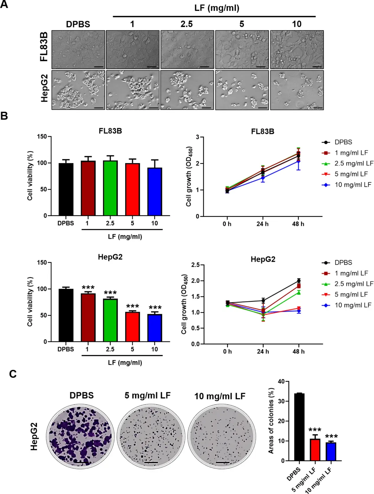
Lactoferrin Exerts Cytotoxic Effects on Human Liver Cancer Cells
Lactoferrin (LF), a natural iron-binding glycoprotein with reported anticancer effects, is investigated for its potential in liver cancer treatment, an area with limited existing studies. To assess the specific cytotoxic effects of LF on liver cancer cells, HepG2, Hep3B, and SK-Hep1 cells were tested, and the results were compared to those of the healthy mouse liver cell line FL83B (Fig. 1). The LF treatments caused considerable inhibition of HepG2 cell proliferation but not healthy mouse FL83B liver cell proliferation (Fig. 1A). Moreover, LF also caused similar inhibitory effects on Hep3B and SK-Hep1 cell proliferation. The LF-induced cytotoxic effects displayed a dose-dependent trend in these cancer cells, especially at concentrations of 5 and 10 mg/ml, which completely inhibited the proliferation of HepG2 and Hep3B cells (Fig. 1B). These results demonstrated that lactoferrin had no significant impact on normal liver cells, even at high doses such as 5 mg/ml and 10 mg/ml, while these doses effectively inhibited cell viability and growth in HCC cell lines (HepG2, Hep3B, and SK-Hep1). Moreover, the LF treatments significantly inhibited the growth of HepG2 and Hep3B cell colonies after an extended incubation time, both in terms of colony size and number (Fig. 1C).
Ask a Question
Write your own review
Description: neuroglial and neuronal character coexpressing ependymoma cell line.
Description: Established by irradiation of the adherent cells in long-term bone marrow cultures derived from C3H/HeNSlc strain mice
Description: Cloned from the C1300 mouse neuroblastoma line. Adapted to culture by alternate in vivo and in vitro passage. Cells have characteristics of mature neurons and secrete the enzymes for the synthesis of ...
Description: EMT6 was established from a transplantable murine mammary carcinoma that arose in a BALB/cCRGL mouse after implantation of a hyperplastic mammary alveolar nodule.

Description: 6-thioguanine resistant mutant of cell line N4 that was established from a mouse of strain A/J with neuroblastoma C1300; cells were described to form increased levels of cAMP in response to ...
- Adipose Tissue-Derived Stem Cells
- Human Neurons
- Mouse Probe
- Whole Chromosome Painting Probes
- Hepatic Cells
- Renal Cells
- In Vitro ADME Kits
- Tissue Microarray
- Tissue Blocks
- Tissue Sections
- FFPE Cell Pellet
- Probe
- Centromere Probes
- Telomere Probes
- Satellite Enumeration Probes
- Subtelomere Specific Probes
- Bacterial Probes
- ISH/FISH Probes
- Exosome Isolation Kit
- Human Adult Stem Cells
- Mouse Stem Cells
- iPSCs
- Mouse Embryonic Stem Cells
- iPSC Differentiation Kits
- Mesenchymal Stem Cells
- Immortalized Human Cells
- Immortalized Murine Cells
- Cell Immortalization Kit
- Adipose Cells
- Cardiac Cells
- Dermal Cells
- Epidermal Cells
- Peripheral Blood Mononuclear Cells
- Umbilical Cord Cells
- Monkey Primary Cells
- Mouse Primary Cells
- Breast Tumor Cells
- Colorectal Tumor Cells
- Esophageal Tumor Cells
- Lung Tumor Cells
- Leukemia/Lymphoma/Myeloma Cells
- Ovarian Tumor Cells
- Pancreatic Tumor Cells
- Mouse Tumor Cells