
Canine Dermal Fibroblasts
Cat.No.: CSC-C9390W
Species: Dog
Source: Dermis; Skin
Morphology: Bipolar
Cell Type: Fibroblast
- Specification
- Background
- Scientific Data
- Q & A
- Customer Review
Canine dermal fibroblasts (CDFs) exhibit a characteristic spindle-shaped, adherent morphology with extensive cytoplasmic processes, forming a confluent monolayer with a swirling, whorled pattern. CDFs are routinely identified by positive immunostaining for vimentin, fibronectin, and collagen type I, while remaining negative for cytokeratin (epithelial marker) and CD31 (endothelial marker).
A fundamental advantage of CDFs lies in their preservation of key in vivo-like functions-including robust synthesis of extracellular matrix components (collagens, elastin, proteoglycans), secretion of growth factors (TGF-β, FGF-2, PDGF), and active participation in wound contraction and re-epithelialization processes. These cells respond to mechanical stimuli, hypoxic conditions, and inflammatory cytokines, mirroring their in-situ behavior. Furthermore, CDFs undergo phenotypic conversion to myofibroblasts upon TGF-β stimulation, characterized by α-smooth muscle actin (αSMA) expression and enhanced contractile activity-a critical process in fibrotic and healing responses.
CDFs are indispensable for preclinical studies of wound healing, hypertrophic scarring, burn injury, dermal drug delivery, and cutaneous toxicology. They are amenable to genetic modification via nucleofection or lentiviral transduction, and compatible with 3D organotypic culture systems. Readily available from commercial sources with rigorous quality control, CDFs serve as a robust, reproducible ex vivo platform bridging veterinary and human dermatological research.
Small Molecules Temporarily Induce Neuronal Features in Adult Canine Dermal Fibroblasts
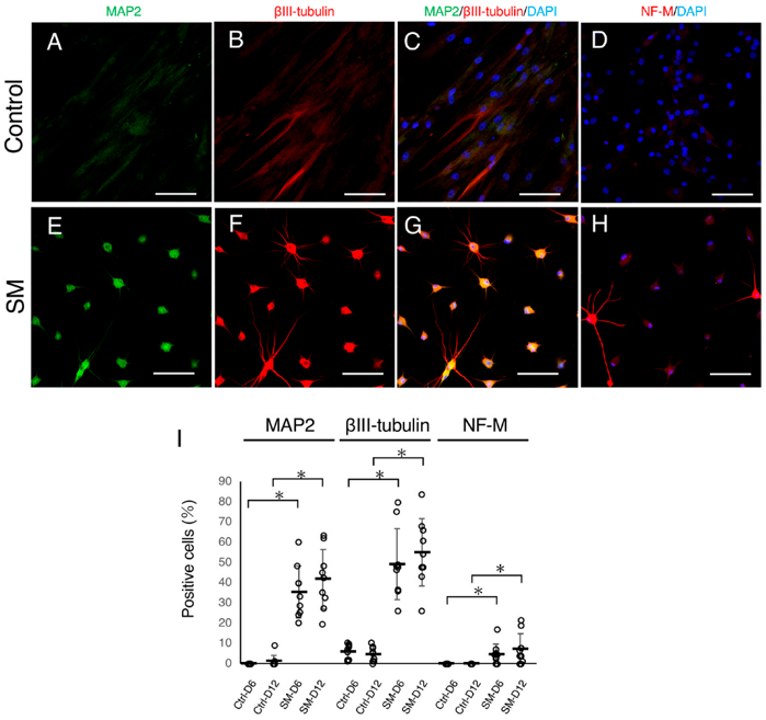
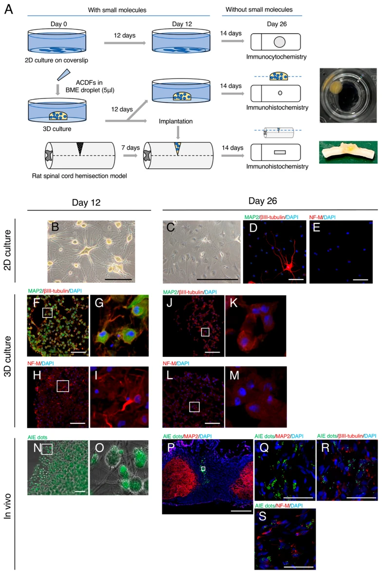
Several methods have been developed to generate neurons from other cell types for performing regeneration therapy and in vitro studies of central nerve disease. Small molecules (SMs) can efficiently induce neuronal features in human and rodent fibroblasts without transgenes. Although canines have been used as a spontaneous disease model of human central nerve, efficient neuronal reprogramming method of canine cells have not been well established. This study aimed to induce neuronal features in adult canine dermal fibroblasts (ACDFs) by SMs and assess the permanency of these changes.
ACDFs treated with eight SMs developed a round-shaped cell body with branching processes and expressed neuronal proteins, including βIII-tubulin, microtubule-associated protein 2 (MAP2), and neurofilament-medium. Transcriptome profiling revealed the upregulation of neuron-related genes, such as SNAP25 and GRIA4, and downregulation of fibroblast-related genes, such as COL12A1 and CCN5. Calcium fluorescent imaging demonstrated an increase in intracellular Ca2+ concentration upon stimulation with glutamate and KCl. Although neuronal features were induced similarly in basement membrane extract droplet culture, they diminished after culturing without SMs or in vivo transplantation into an injured spinal cord. In conclusion, SMs temporarily induce neuronal features in ACDFs. However, the analysis of bottlenecks in the neuronal induction is crucial for optimizing the process.
Ask a Question
Write your own review
Description: Dog Liver Endothelial Cells from Creative Bioarray are isolated from tissue of dog liver. Dog Liver Endothelial Cells are grown in T25 tissue culture flasks pre-coated with gelatin-based coating ...
Description: Canine Astrocytes from Creative Bioarray are isolated from canine brain tissue. The method we use to isolate canine astrocytes were developed based on a combination of established and our proprietary ...
Description: Canine Mammary Microvascular Endothelial Cells from Creative Bioarray are isolated from breast of pathogen-free laboratory Canine. Canine Mammary Microvascular Endothelial Cells are grown in T25 ...
Description: Canine Chondrocytes (CnC) provided by Creative Bioarray are isolated from normal canine articular cartilage tissue. The cells are frozen at passage 1 and each vial contains at least 0.5*10^6 cells. ...
Description: Canine Pancreatic Microvascular Endothelial Cells from Creative Bioarray are isolated from Pancreatic Microvascular of pathogen-free laboratory Canine. Canine Pancreatic Microvascular Endothelial ...
Description: Canine Prostate Microvascular Endothelial Cells from Creative Bioarray are isolated from prostate of pathogen-free laboratory Canine. Canine Prostate Microvascular Endothelial Cells are grown in T25 ...