
Strain Balb/c Mouse Mesenchymal Stem Cells
- Specification
- Background
- Scientific Data
- Q & A
- Customer Review
Balb/c Mouse Mesenchymal Stem Cells (MSCs) are primary, multipotent stromal cells isolated from the bone marrow of the inbred Balb/c mouse strain. They exhibit the classical fibroblast-like, spindle-shaped morphology and adhere firmly to plastic surfaces. These cells are defined by their specific immunophenotype, uniformly expressing mesenchymal markers such as CD29, CD44, CD73, and Sca-1, while lacking hematopoietic lineage markers like CD11b, CD34, and CD45. Functionally, they possess robust trilineage differentiation potential, capable of differentiating into osteoblasts, adipocytes, and chondrocytes under specific inductive conditions.
Culturally, they are maintained in a nutrient-rich basal medium supplemented with serum and growth factors under standard conditions (37°C, 5% CO₂). Due to the genetic homogeneity of the Balb/c background, these MSCs offer high experimental reproducibility. They are widely utilized in tissue engineering, immunomodulation studies, and as a preferred model for in vivo homing assays, particularly in Balb/c-based xenograft or syngeneic transplantation systems.
Adipose-Derived Mesenchymal Stem Cells Attenuate Cutaneous Leishmaniasis Progression in BALB/c Mice
Leishmaniasis remains a major public health challenge, necessitating immunotherapies that activate macrophages and promote TH1 responses. While mesenchymal stem cells (MSCs) exhibit immunomodulatory effects, this study evaluated the impact of adipose-derived MSCs (AD-MSCs) on cutaneous leishmaniasis in BALB/c mice.
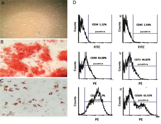
AD-MSCs displayed a fibroblast-like morphology and successfully differentiated into osteocytes and adipocytes (Fig. 1A-C). Immunophenotyping confirmed high expression of MSC markers (CD90: 44.68%, CD105: 61.55%, CD73: 46.82%, CD29: 95.7%) and low expression of hematopoietic markers (CD45: 1.54%, CD34: 1.12%) (Fig. 1D).
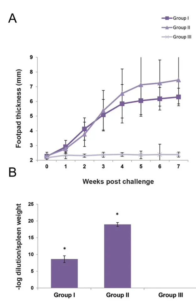
Following L. majorinfection, disease progression was monitored for 7 weeks. Infected control mice (Group II) developed lesions by week 2, with footpad thickness increasing significantly from weeks 3 to 7, culminating in necrosis. In contrast, AD-MSC-treated mice (Group I) exhibited delayed lesion formation with reduced severity. Footpad thickness in the treated group was significantly lower than in controls from weeks 4 to 7 (Fig. 2A). These findings demonstrate that AD-MSC administration attenuates disease progression in experimental cutaneous leishmaniasis.
Ask a Question
Write your own review
- Adipose Tissue-Derived Stem Cells
- Human Neurons
- Mouse Probe
- Whole Chromosome Painting Probes
- Hepatic Cells
- Renal Cells
- In Vitro ADME Kits
- Tissue Microarray
- Tissue Blocks
- Tissue Sections
- FFPE Cell Pellet
- Probe
- Centromere Probes
- Telomere Probes
- Satellite Enumeration Probes
- Subtelomere Specific Probes
- Bacterial Probes
- ISH/FISH Probes
- Exosome Isolation Kit
- Human Adult Stem Cells
- Mouse Stem Cells
- iPSCs
- Mouse Embryonic Stem Cells
- iPSC Differentiation Kits
- Mesenchymal Stem Cells
- Immortalized Human Cells
- Immortalized Murine Cells
- Cell Immortalization Kit
- Adipose Cells
- Cardiac Cells
- Dermal Cells
- Epidermal Cells
- Peripheral Blood Mononuclear Cells
- Umbilical Cord Cells
- Monkey Primary Cells
- Mouse Primary Cells
- Breast Tumor Cells
- Colorectal Tumor Cells
- Esophageal Tumor Cells
- Lung Tumor Cells
- Leukemia/Lymphoma/Myeloma Cells
- Ovarian Tumor Cells
- Pancreatic Tumor Cells
- Mouse Tumor Cells