
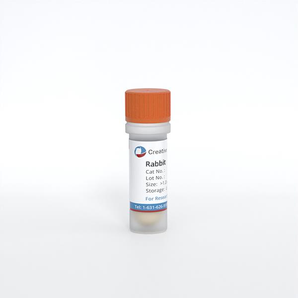
Rabbit Tenocytes
Cat.No.: CSC-C5165S
Species: Rabbit
Source: Tendon
Cell Type: Tenocyte
- Specification
- Background
- Scientific Data
- Q & A
- Customer Review
Rabbit tenocytes from Creative Bioarray are isolated from the rabbit tendon tissue. The method we use to isolate rabbit tenocytes was developed based on a combination of established and our proprietary methods. The rabbit tenocytes from Creative Bioarray are characterized by immunofluorescence with antibodies specific to type I collagen. Each vial contains 0.5x10^6 cells per ml and is delivered frozen.
Rabbit tenocytes are primary fibroblast-like cells derived from rabbit tendons, most typically from Achilles or flexor tendons. They are the major cellular component of tendon tissue and produce and maintain the dense extracellular matrix that is rich in type I collagen, providing the mechanical strength and structural integrity necessary to transmit loads.
Rabbit tenocytes in vitro show a characteristic elongated spindle-shaped appearance and grow as adherent monolayers. They are commonly cultured in a basal medium enriched with nutrients and supplemented with serum under conventional conditions (37 °C, 5% CO2). Cultures must be carefully controlled to avoid undesired differentiation or loss of phenotypic. Overpassage can lead to dedifferentiation and decreased collagen synthesis.
These cells represent an important model to research the biology of tendons and the mechanisms of tendon healing as well as pathological problems such as tendinopathy and adhesive scarring due to the near biomechanical resemblance between rabbit and human tendons. They are widely applied in tissue engineering to investigate scaffold biocompatibility, to evaluate growth factor therapy and to create regenerative techniques for tendon repair; thus, they provide an important pre-clinical platform between in vitro studies and clinical applications.
Characterization of Rabbit ADSCs and Tenocytes for Cell-Free Tendon Therapy Development
Tendon injuries exhibit poor healing due to the tissue's hypovascular and hypocellular nature, necessitating advanced therapies. To develop a cell-free strategy, Wolint et al. analyzed the secretomes of rabbit adipose-derived stem cells (rbADSCs) and an rbADSC-tenocyte co-culture using LC-MS/MS.
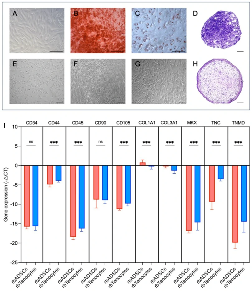
rbADSCs were isolated from New Zealand white rabbits (Fig. 1A). Passage 1 cells demonstrated multipotent differentiation potential into adipogenic, osteogenic, and chondrogenic lineages, confirmed by Alizarin Red, Oil Red-O, and Toluidine Blue staining (Fig. 1B-D, F-H). Both rbADSCs and rabbit tenocytes (rbTenocytes) were characterized for surface marker expression (Fig. 1E). rbADSCs exhibited high expression of MSC markers (CD44, CD90, CD105) and extracellular matrix genes (COL I, COL III, TNC), with low expression of hematopoietic markers (CD34, CD45) and tenogenic markers (MKX, TNMD) (Fig. 1I). Conversely, rbTenocytes showed significant upregulation of CD44, CD45, MKX, TNC, and TNMD compared to rbADSCs (Fig. 1I). These results confirm the expected MSC phenotype for rbADSCs and the tendon-specific genotype for rbTenocytes.
Ask a Question
Write your own review

Description: Rabbit Hepatocytes are derived from the liver of New Zealand White Rabbit.
Description: The aortic arch is the top part of the main artery carrying blood away from the heart. It is the connection between the ascending and descending aorta, and its central part is formed by the left 4th ...
Description: The synovium secretes synovial fluid, which plays an important role in joint movement. The normal synovium has two layers, a thin cellular layer (luminal layer) and a vascular layer (subintima). ...

Description: The pituitary gland is an important endocrine gland in the body that secretes growth hormone and adrenocorticotropic hormone. It plays an important role in the growth and development of the body, ...
Description: The oral epthelial cells are responsible for important functions, like the primary protection of oral mucosa against external aggressions building a mechanical barrier against microorganisms, ...
Description: The carotid arteries are major blood vessels in the neck that supply blood to the brain, neck, and face. There are two carotid arteries, one on the right and one on the left. In the neck, each ...