
Immortalized Porcine Alveolar Macrophages
- Specification
- Background
- Scientific Data
- Q & A
- Customer Review
Never can cryopreserved cells be kept at -20 °C
Porcine alveolar macrophages (PAMs) are a key component of the pulmonary immune system, primarily residing in the alveolar space to eliminate inhaled pathogens through phagocytosis. The Immortalized Porcine Alveolar Macrophages (iPAMs) cell line maintains macrophage-like morphology featuring a wrinkled membrane surface with cellular extensions. Upon exposure to pathogens or LPS iPAMs generate multiple pro-inflammatory cytokines including IL-1β, TNF-α, IL-6 and IL-12B. These cytokines play crucial roles in starting immune system reactions. iPAMs represent the main cellular target for the porcine reproductive and respiratory syndrome virus (PRRSV). During infection iPAMs mount antiviral defenses via ISGylation and block viral replication by engulfing and destroying viral particles.
iPAMs also possess immune regulatory functions. The immune functions of these cells change according to their polarization status which comprises M1 macrophages that exhibit pro-inflammatory characteristics and M2 macrophages that perform anti-inflammatory roles throughout various immune response phases. Through the secretion of chemokines CCL2 and CXCL10 iPAMs can recruit additional immune cells to their location. Since PAMs represent the main cellular targets for PRRSV infection iPAMs provide crucial insights for studies on PRRSV infection patterns replication processes and immune system evasion. Scientists use iPAMs to create laboratory-based porcine respiratory disease models to study disease development and assess treatment methods.
Modulation of Swine Influenza A Virus Replication in Co-Infected PCV2b/SwIV H1N1 Cells
The porcine respiratory disease complex (PRDC) is a significant economic burden in the swine industry, with co-infections involving porcine circovirus type 2 (PCV2) and swine influenza A virus (SwIV) playing a pivotal role. PCV2, a single-stranded DNA virus, complicates swine health by altering immune responses, facilitating co-infection with other pathogens like SwIV, an RNA virus prevalent in global swine populations.
Burgher et al. examined the co-infection pathogenesis between PCV2b and SwIV at the cellular level through studies on tracheal epithelial cells and alveolar macrophages within porcine respiratory tissue. The immunofluorescence assay (IFA) demonstrated simultaneous presence of PCV2b and SwIV H1N1 antigens in cells from co-infected newborn pig tracheal epithelial cell line (NPTr) and immortalized porcine alveolar macrophage (iPAM 3D4/21) (Fig. 1). Similar results were noted for PCV2b/SwIV H3N2 co-infections. PCV2b internalized efficiently in both cell types, as indicated by similar percentages of PCV2b-positive cells. However, more SwIV H1N1-positive cells appeared in NPTr cells than in iPAM 3D4/21 cells. SwIV H1N1/H3N2 infectious titers and PCV2b genome quantities were compared in co-infected cells versus single-infected cells at various times post-infection (Fig. 2). In single-infected NPTr cells, SwIV H1N1/H3N2 titers at 24 hpi were higher than in iPAM 3D4/21 cells, consistent with SwIV's preference for respiratory epithelial cells. PCV2b qPCR results were similar in both cell types (Fig. 3). PCV2b modulated SwIV replication kinetics; in NPTr cells, SwIV titers decreased significantly at 24 hpi in co-infected cells versus SwIV-only cells (Fig. 2A and C). Conversely, in iPAM 3D4/21 cells, SwIV titers were higher in co-infected than single-infected cells (Fig. 2B and D), though SwIV did not impact PCV2b replication during co-infection (Fig. 3).
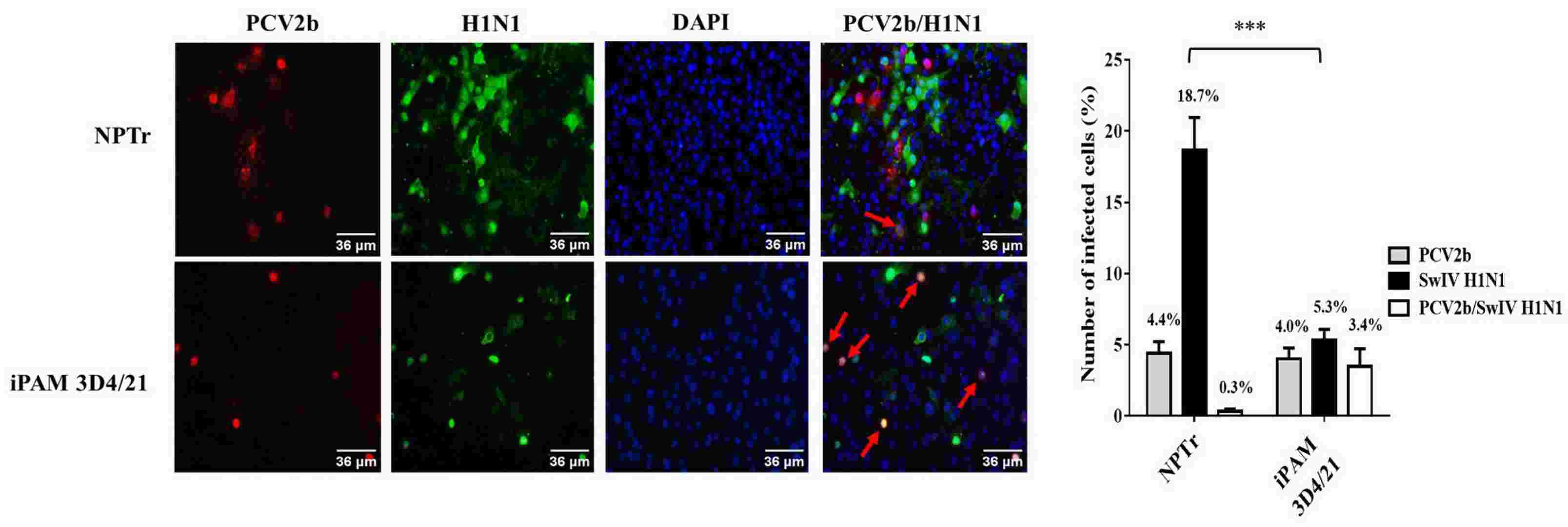 Fig. 1. PCV2b and SwIV H1N1 co-localization in both NPTr and iPAM 3D4/21 co-infected cells (Burgher Pulgaron Y, Provost C, et al., 2023).
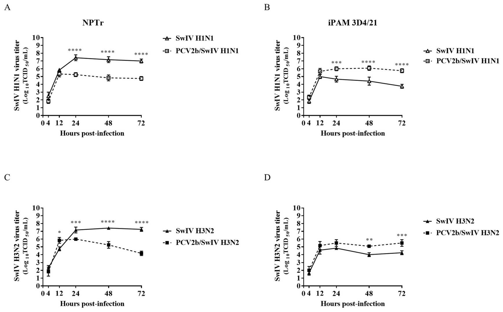 Fig. 2. PCV2b co-infection effects on SwIV replication in infected NPTr and iPAM 3D4/21 cells (Burgher Pulgaron Y, Provost C, et al., 2023).
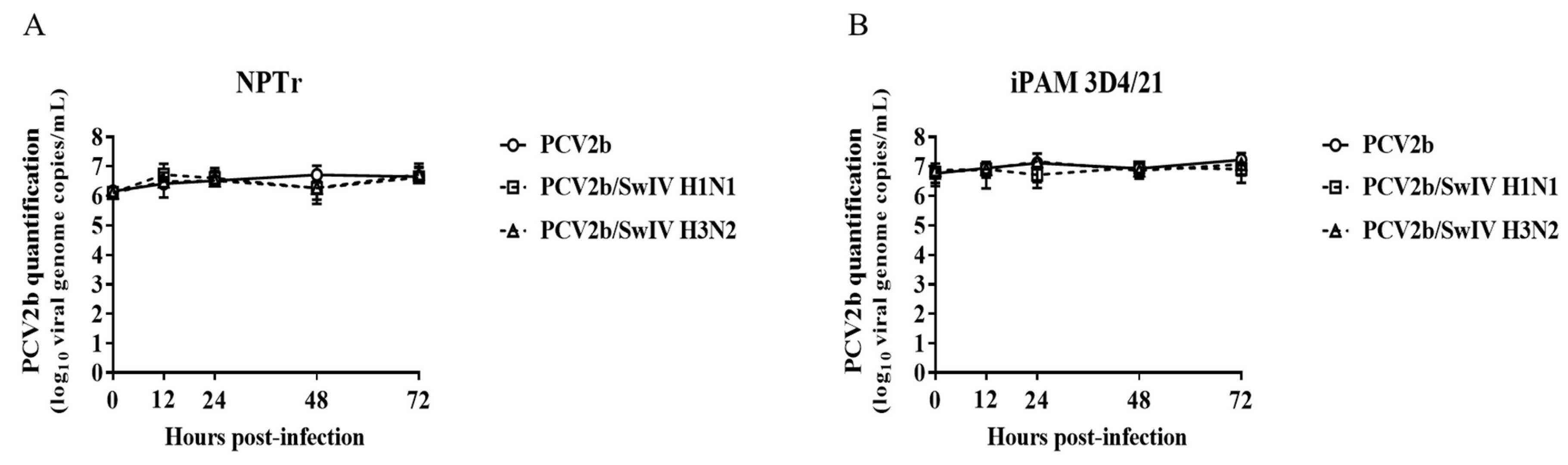 Fig. 3. SwIV co-infection effects on PCV2b replication in infected NPTr and iPAM 3D4/21 cells (Burgher Pulgaron Y, Provost C, et al., 2023).
SVA Exerts Cytotoxicity to 3D4/21 Cells, and to a Greater Extent to IBRS-2 Cells
SVA is an emerging RNA virus affecting pigs. Though adaptive immune responses have been explored, comprehensive insights into SVA-induced innate immunity are scarce due to inadequate cell culture models. Since SVA is a naturally occurring oncolytic virus, Dang' s team speculated that SVA could lyse immortalized porcine alveolar macrophage (iPAM 3D4/21) cells if they are permissive to SVA replication. In the following experiments, in parallel, IBRS-2 cells were used as a telling control due to its high susceptibility to SVA replication. RAW 264.7 cells were also included as its resistance to SVA challenge. Infection with SVA induced clear CPE in 3D4/21 cells, as cells became rounded, ruptured and partially detached from the culture flask (Fig. 4A). Assessment of the degree of CPE demonstrated that SVA infection led to the loss of cell viability of IBRS-2 and 3D4/21 cells, whereas it had no major effect on RAW 264.7. Of note, same amount of SVA inoculum resulted in a more pronounced cell killing in IBRS-2 cells (Fig. 4B). Consistently, Annexin-V/PI staining showed that SVA induced more evident apoptosis in IBRS-2 than 3D4/21 cells, especially late apoptosis (ANXV+PI+), irrespective of incubation time, mirroring the observations in Fig. 4B. Collectively, SVA exhibited cytotoxicity to 3D4/21 cells, and to a greater extent to IBRS-2 cells.
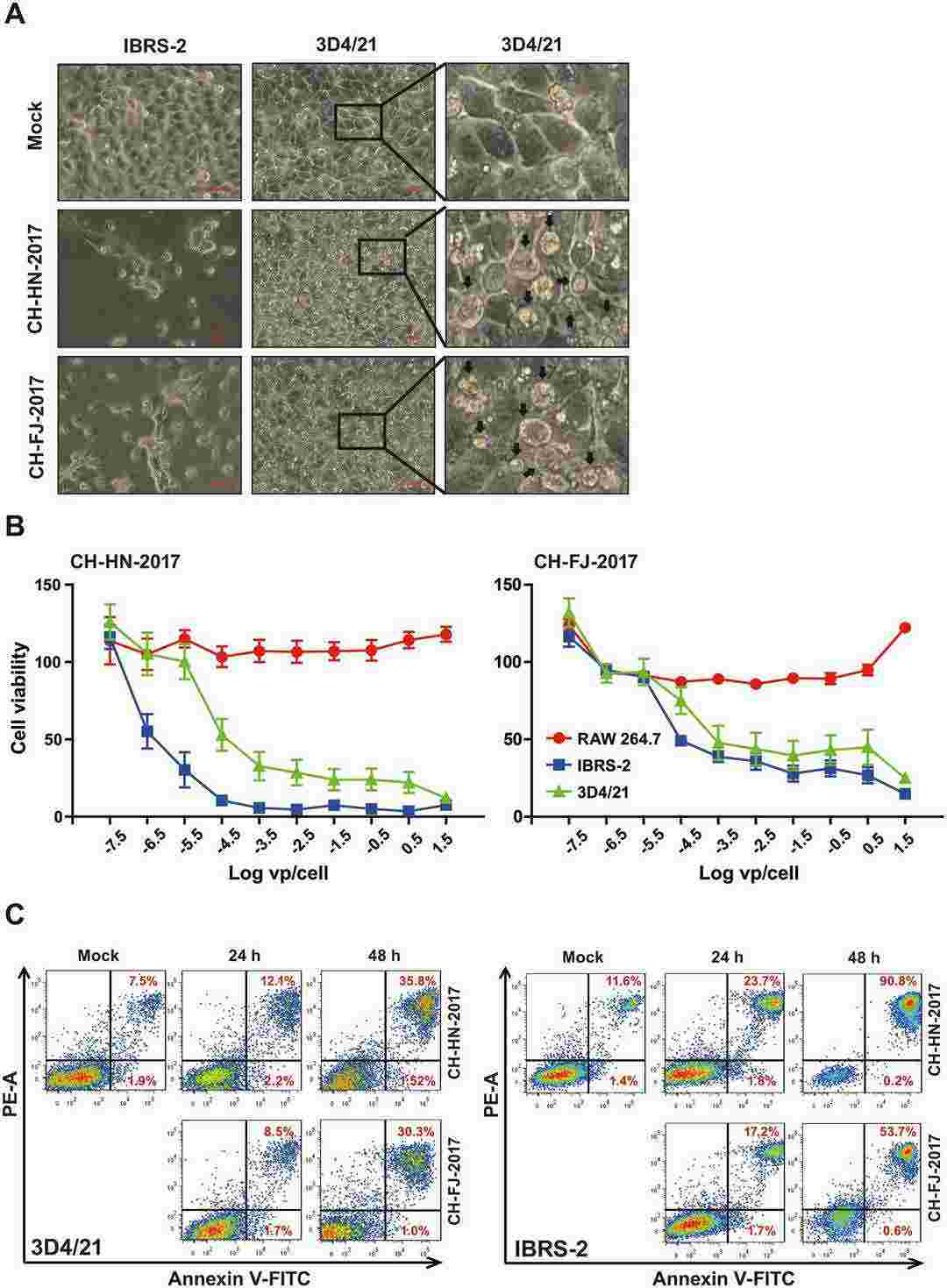 Fig. 4. Cytotoxicity of SVA strains CH–HN-2017 and CH-FJ-2017 to 3D4/21 cells (Dang W, Li T, et al., 2023).
Ask a Question
Write your own review
- Adipose Tissue-Derived Stem Cells
- Human Neurons
- Mouse Probe
- Whole Chromosome Painting Probes
- Hepatic Cells
- Renal Cells
- In Vitro ADME Kits
- Tissue Microarray
- Tissue Blocks
- Tissue Sections
- FFPE Cell Pellet
- Probe
- Centromere Probes
- Telomere Probes
- Satellite Enumeration Probes
- Subtelomere Specific Probes
- Bacterial Probes
- ISH/FISH Probes
- Exosome Isolation Kit
- Human Adult Stem Cells
- Mouse Stem Cells
- iPSCs
- Mouse Embryonic Stem Cells
- iPSC Differentiation Kits
- Mesenchymal Stem Cells
- Immortalized Human Cells
- Immortalized Murine Cells
- Cell Immortalization Kit
- Adipose Cells
- Cardiac Cells
- Dermal Cells
- Epidermal Cells
- Peripheral Blood Mononuclear Cells
- Umbilical Cord Cells
- Monkey Primary Cells
- Mouse Primary Cells
- Breast Tumor Cells
- Colorectal Tumor Cells
- Esophageal Tumor Cells
- Lung Tumor Cells
- Leukemia/Lymphoma/Myeloma Cells
- Ovarian Tumor Cells
- Pancreatic Tumor Cells
- Mouse Tumor Cells