
Cynomolgus Monkey Kidney Fibroblasts
Cat.No.: CSC-C4725L
Species: Monkey
Source: Kidney
Cell Type: Fibroblast
- Specification
- Background
- Scientific Data
- Q & A
- Customer Review
Never can cryopreserved cells be kept at -20 °C.
Cynomolgus Monkey Kidney Fibroblasts are primary fibroblast cells originally derived from kidney tissue of cynomolgus monkeys. The cells phenotypically resemble renal fibroblasts in vivo and serve as a non-human primate in vitro model commonly employed for biomedical and translational research applications. Being closely related to humans, cynomolgus monkey kidney fibroblasts allow for research with high translational relevance between preclinical and clinical development.
Cynomolgus monkey kidney fibroblasts have a classic spindle-shaped fibroblast-like morphology and will grow as an adherent monolayer when maintained in standard cell culture conditions. Functionally, they have been shown to contribute to ECM production, tissue remodeling, and stromal-epithelial communication. Cynomolgus monkey kidney fibroblasts express expected fibroblast markers and have been shown to react strongly to profibrotic treatments like TGF-β. Because of these properties, the cells are often used to study mechanisms of renal fibrosis and disease.
These cells are commonly applied in nephrotoxicity assessment, drug metabolism and safety studies, and fibrosis-related signaling pathway analysis. In addition, they are useful for evaluating species-specific drug responses and off-target effects in non-human primate systems. The cells have also been used for characterization of species-specific responses to drugs and off-target drug effects in non-human primates.
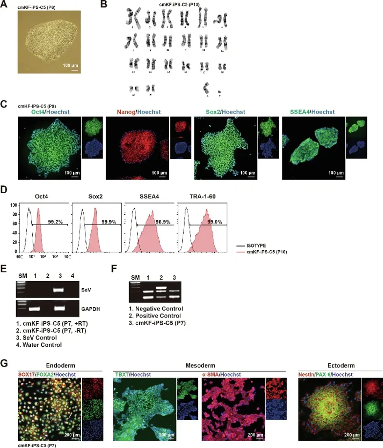
Generation and Characterization of Cynomolgus Monkey Kidney Fibroblasts (cmKF)-Derived Induced Pluripotent Stem Cells (cmKF-iPS-C5)
Cynomolgus monkeys are genetically and physiologically closer to humans than other mammals, making them prime candidates for developmental and biomedical studies. In this study, Zhen's team reprogrammed cynomolgus monkey kidney fibroblasts (cmKFs) into iPSCs using Reprogramming Kit (Oct3/4, Sox2, KLF4, c-Myc) as negative controls for their autologous transplantations.
Colony formed displayed embryonic stem cell-like morphology and high nucleus-to-cytoplasmic ratios (Fig. 1A). STR analyses matched parental fibroblasts, and G-banding showed normal male karyotype (20, XY) (Fig. 1B). Immunostaining and flow cytometry confirmed the expression of pluripotency markers including core transcription factors (Oct4, NANOG, SOX2) and surface antigens (TRA-1-60, SSEA4) (Fig. 1C, D). PCR confirmed clearance of Sendai virus reprogramming factors and absence of Mycoplasma infection (Fig. 1E, F). Functional pluripotency assays showed differentiation along the three germ layers of endoderm (FOXA2 and SOX17), mesoderm (TBXT and α-SMA), and ectoderm (Nestin and PAX-6) (Fig. 1G).
Ask a Question
Write your own review
Description: Cynomolgus Monkey Primary Spleen Fibroblasts are isolated from Cynomolgus Cynomolgus Monkey spleen tissue.
Description: Cynomolgues Monkey Pancreatic Islets from Creative Bioarray are isolated from the Cynomolgus Monkey pancreas using Collagenase P and purified using Ficoll density gradient.
Description: Cynomolgus Monkey Pancreatic Epithelial Cells from Creative Bioarray are isolated from normal Cynomolgus Monkey pancreatic tissue. Cynomolgus Monkey Pancreatic Epithelial Cells are grown in T25 ...
Description: Cynomolgus Monkey Primary Bladder Epithelial Cells are isolated from normal Cynomolgus Monkey bladder tissue.
Description: Cynomolgus Monkey Dermal Lymphatic Endothelial Cells from Creative Bioarray are isolated from Cynomolgus monkey skin tissue. Monkey Dermal Lymphatic Endothelial Cells are grown in T25 tissue culture ...
Description: Cynomolgus Monkey Proximal Tubular Epithelial Cells from Creative Bioarray are isolated from normal Cynomolgus Monkey proximal tubular. Cynomolgus Monkey Proximal Tubular Epithelial Cells are grown ...