Rabbit Aortic Endothelial Cells
Cat.No.: CSC-C4199X
Species: Rabbit
Source: Aorta
Cell Type: Endothelial Cell
- Specification
- Background
- Scientific Data
- Q & A
- Customer Review
Rabbit aortic endothelial cells (RAECs) are primary endothelial cells isolated from the tunica intima of the rabbit aorta. They form a confluent, cobblestone monolayer and express hallmark endothelial markers including von Willebrand factor (vWF), CD31 (PECAM-1), and VE-cadherin, while exhibiting uptake of acetylated low-density lipoprotein (Ac-LDL) and tube formation capacity on Matrigel.
The rabbit model has been historically and continues to be a cornerstone in cardiovascular research due to its anatomical and physiological similarities to the human vasculature, particularly in terms of vessel size, hemodynamic profiles, and lipoprotein metabolism. Unlike mice, rabbits are susceptible to diet-induced hypercholesterolemia and develop atherosclerotic lesions recapitulating human pathology, making RAECs highly relevant for translational studies.
Key advantages of RAECs include: (1) well-characterized responsiveness to shear stress, inflammatory cytokines (e.g., TNF-α, IL-1β), and oxidized LDL, enabling investigation of endothelial activation and dysfunction; (2) ready isolation from relatively large vessels, yielding sufficient cell numbers for passaging and experimental replicates; (3) preservation of in vivo-like properties over limited passages, including expression of eNOS and responsiveness to acetylcholine, which are often lost in immortalized lines; (4) cost-effectiveness and availability compared to human aortic endothelial cells (HAECs); (5) historical comparability - extensive literature on rabbit endothelium provides validated protocols and benchmarks.
Co-transplantation of Angiotensin II-Pretreated Mesenchymal Stem Cells and Endothelial Cells in Early Steroid-Induced Osteonecrosis of the Femoral Head
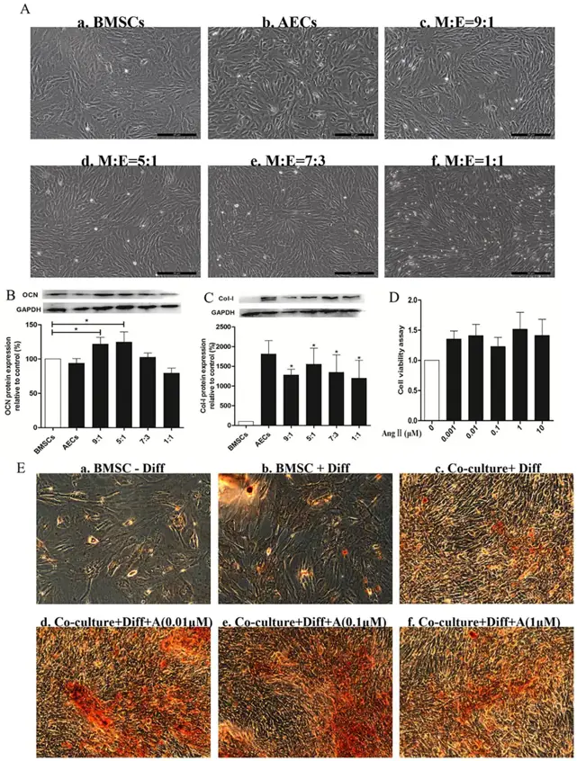
Although mesenchymal stem cells (MSCs) and endothelial cells (ECs) co-culture enhancing proliferation and osteogenic differentiation of MSCs and form more mature vasculature in vivo, it remains unknown whether the co-culture cells are able to repair osteonecrosis of the femoral head (ONFH). In this study, we explored the roles and mechanisms of co-transplantation of angiotensin II (Ang II)-MSCs and ECs in repairing early ONFH.
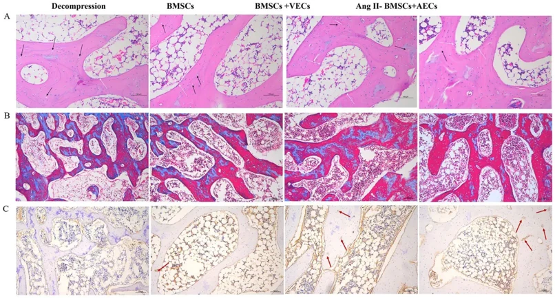
In vitro, when MSCs and ECs were co-cultured in a ratio of 5:1, both types of cells managed to proliferate and induce both osteogenesis and angiogenesis. Then, we established a rabbit model of steroid-induced ONFH and co-transplantation of Ang II-MSCs and ECs through the tunnel of core decompression. Four weeks later, histological and Western blot analyses revealed that ONFH treated with Ang II-MSCs and ECs may promote ossification and revascularization by increasing the expression of collagen type I, runt-related transcription factor 2, osteocalcin, and vascular endothelial growth factor in the femoral head. Our data suggest that co-transplantation of Ang II-MSCs and ECs was able to rescue the early steroid-induced ONFH via promoting osteogenesis and angiogenesis, which may be regarded as a novel therapy for the treatment of ONFH in a clinical setting.
Ask a Question
Write your own review

Description: Rabbit Hepatocytes are derived from the liver of New Zealand White Rabbit.
Description: The aortic arch is the top part of the main artery carrying blood away from the heart. It is the connection between the ascending and descending aorta, and its central part is formed by the left 4th ...

Description: The synovium secretes synovial fluid, which plays an important role in joint movement. The normal synovium has two layers, a thin cellular layer (luminal layer) and a vascular layer (subintima). ...
Description: The pituitary gland is an important endocrine gland in the body that secretes growth hormone and adrenocorticotropic hormone. It plays an important role in the growth and development of the body, ...
Description: The oral epthelial cells are responsible for important functions, like the primary protection of oral mucosa against external aggressions building a mechanical barrier against microorganisms, ...
Description: The carotid arteries are major blood vessels in the neck that supply blood to the brain, neck, and face. There are two carotid arteries, one on the right and one on the left. In the neck, each ...