
QualiCell® Human Neuronal Stem Cells (Hippocampus) -XLC418
- Specification
- Background
- Scientific Data
- Q & A
- Customer Review
Human hippocampal neural stem cells (NSCs) reside in the subgranular zone (SGZ) of the dentate gyrus (DG), one of the two principal neurogenic niches in the adult mammalian brain. Unlike cortical NSCs, which are primarily fetal-derived and generate glutamatergic projection neurons, hippocampal NSCs sustain lifelong neurogenesis, continuously producing new granule neurons that integrate into existing hippocampal circuits. This enduring plasticity underpins their unique advantages for both fundamental neuroscience and translational applications.
First, lifelong neurogenic capacity – hippocampal NSCs remain active throughout life, generating neurons that critically support learning, memory, and pattern separation. This property is absent in cortical NSCs, which exhibit limited postnatal neurogenesis.
Second, region-specific neuronal output – upon differentiation, hippocampal NSCs preferentially generate DG granule neurons—a restricted lineage distinct from the diverse pyramidal and interneuron subtypes produced by cortical NSCs. This specified fate makes them uniquely suited for modelling hippocampal-dependent cognitive processes and disorders.
Third, therapeutic relevance for memory-related pathologies – hippocampal NSCs are directly implicated in Alzheimer's disease, epilepsy, depression, and age-related cognitive decline. Transplantation of human hippocampal NSCs has been shown to rescue cognitive deficits and reinforce synaptic networks in preclinical models.
Fourth, human-specific relevance – unlike rodent models, human hippocampal NSCs exhibit deeper quiescence and lower baseline neurogenesis, more accurately reflecting the human condition. Single-cell studies have revealed molecular heterogeneity—quiescent, primed, and active NSC states—providing a framework for understanding age-related decline and disease vulnerability.
The Effects of Cannabidiol and Its Main Metabolites on Human Neural Stem Cells
In the present study, human neural stem cells (NSCs) were treated with cannabidiol (CBD) and its metabolites (7-OH-CBD and 7-COOH-CBD) to understand how the drug may affect fetal brain development. NSCs were also treated with delta-9 tetrahydrocannabinol (THC) for comparison purposes.
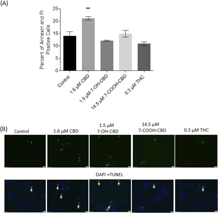
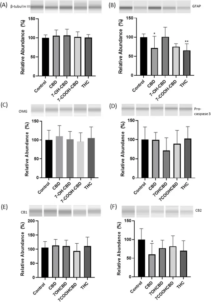
CBD, 7-OH-CBD and 7-COOH-CBD dose-dependently reduced NSC viability. CBD and 7-OH-CBD reduced NSC number at the G1 phase. A 24 h exposure did not cause significant change in NSC proliferation. At concentrations comparable to those detected in human blood, longer exposures to CBD, 7-OH-CBD and 7-COOH-CBD caused more obvious cell death. After NSCs differentiation, CBD treatment reduced GFAP and cannabinoid receptor 2 (CB2) expression. THC treatment reduced the GFAP expression, but the change in CB2 expression did not reach statistical significance. The expression of cannabinoid receptor 1 (CB1) and beta-tubulin III were not significantly altered by drug exposures. The study demonstrated that clinically relevant concentrations of CBD, 7-OH-CBD and 7-COOH-CBD affect basic physiological features of human NSCs. After NSC differentiation, the reduced expression of CB2 receptors and GFAP on differentiated cells further indicated the vulnerability of developing central nervous system to CBD and THC. These data will help to contextualize in vivo neurodevelopmental studies that may not accurately model human metabolite profiles of CBD.
Ask a Question
Write your own review
- You May Also Need
- Adipose Tissue-Derived Stem Cells
- Human Neurons
- Mouse Probe
- Whole Chromosome Painting Probes
- Hepatic Cells
- Renal Cells
- In Vitro ADME Kits
- Tissue Microarray
- Tissue Blocks
- Tissue Sections
- FFPE Cell Pellet
- Probe
- Centromere Probes
- Telomere Probes
- Satellite Enumeration Probes
- Subtelomere Specific Probes
- Bacterial Probes
- ISH/FISH Probes
- Exosome Isolation Kit
- Human Adult Stem Cells
- Mouse Stem Cells
- iPSCs
- Mouse Embryonic Stem Cells
- iPSC Differentiation Kits
- Mesenchymal Stem Cells
- Immortalized Human Cells
- Immortalized Murine Cells
- Cell Immortalization Kit
- Adipose Cells
- Cardiac Cells
- Dermal Cells
- Epidermal Cells
- Peripheral Blood Mononuclear Cells
- Umbilical Cord Cells
- Monkey Primary Cells
- Mouse Primary Cells
- Breast Tumor Cells
- Colorectal Tumor Cells
- Esophageal Tumor Cells
- Lung Tumor Cells
- Leukemia/Lymphoma/Myeloma Cells
- Ovarian Tumor Cells
- Pancreatic Tumor Cells
- Mouse Tumor Cells