
Immortalized Human Astrocytes, fetal-hTERT
Cat.No.: CSC-I9064L
Species: Homo sapiens
Source: Brain
Morphology: Multipolar
Culture Properties: Adherent
- Specification
- Background
- Scientific Data
- Q & A
- Customer Review
Note: Never can cells be kept at -20 °C.
CIK-HT013 HT® Lenti-hTERT Immortalization Kit
Immortalized Human Astrocytes, were derived from human fetal brain and transduced with lentivirus encoding human telomerase reverse transcriptase (hTERT). Restoring telomerase activity allows these cells to bypass replicative senescence, while maintaining a normal diploid karyotype and physiologic morphology for hundreds of population doublings. Compared to primary astrocytes which senesce within days to weeks of in vitro culture, fetal‑hTERT cells allow for a stable, scalable, renewable source of cells which exhibit many fundamental characteristics of astrocytes such as GFAP expression, cytokine secretion, and glutamate uptake.
Applications of this cell line go beyond serving as a "generic" model of glia in neurobiology. Their use includes serving as an in vitro model for isolating mechanisms of the aging epigenome by decoupling replication-driven DNA methylation (DNAm) drift from age itself. Additionally, they can be used to study the blood‑brain barrier (BBB) and neuroinflammation, as they can be cocultured with endothelial cells to better replicate in vitro models of the BBB. In translational science, these cells have been used to understand viral neurotropism (SARS-CoV-2, Zika virus) and screen for neuroprotective drugs. Strictly research use (BSL-2).
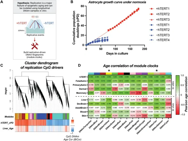
Modeling a Shared Replication-Associated Epigenetic Signature in Aging and Cancer
Aging is a primary cancer risk factor, a connection often linked to accumulating somatic mutations, though this is likely an incomplete explanation. Minteer et al. identified a common epigenetic replication signature between aging and cancer, which they modeled using DNA methylation (DNAm) from extensively passaged, immortalized human cells and validated in clinical tissues.
They longitudinally profiled DNAm in serially passaged, hTERT-immortalized fetal astrocytes, chosen for their minimal fitness selection in culture compared to adult cells and the hypothesized cell type-independence of replication-driven DNAm changes. These cells serve as a physiologically relevant glial model with enhanced proliferative capacity. Their lifespan was extended by over 700% compared to non-immortalized astrocytes. After more than 73 cumulative population doublings (cPDs), cells exhibited no signs of growth arrest, genomic instability, or telomere erosion, enabling isolation of replication-associated epigenetic changes (Fig. 1A and B).
Ask a Question
Write your own review
Description: Nasal epithelial cells form the outermost protective layer against environmental factors. They clean, humidify, and warm inhaled air. They produces mucus, which binds particles that are subsequently ...
Description: Immortalized Human Splenic Endothelial Cells-SV40 have been obtained immortalizing Human Splenic Endothelial Cells with SV40LT expressing lentiviral particles. Immortalized cells were controlled ...
Description: Immortalized Human Corneal Epithelial Cells-SV40 have been obtained immortalizing Human Corneal Epithelial Cells with Lenti-SV40 Lentivirus. Immortalized cells were controlled passaging side by side ...

Description: Immortalized Human Lymphatic Endothelial Cells-SV40 were developed from human tissues transduced with a lentiviral expression vector containing the SV40T gene. The cell line was continuously cultured ...
- Adipose Tissue-Derived Stem Cells
- Human Neurons
- Mouse Probe
- Whole Chromosome Painting Probes
- Hepatic Cells
- Renal Cells
- In Vitro ADME Kits
- Tissue Microarray
- Tissue Blocks
- Tissue Sections
- FFPE Cell Pellet
- Probe
- Centromere Probes
- Telomere Probes
- Satellite Enumeration Probes
- Subtelomere Specific Probes
- Bacterial Probes
- ISH/FISH Probes
- Exosome Isolation Kit
- Human Adult Stem Cells
- Mouse Stem Cells
- iPSCs
- Mouse Embryonic Stem Cells
- iPSC Differentiation Kits
- Mesenchymal Stem Cells
- Immortalized Human Cells
- Immortalized Murine Cells
- Cell Immortalization Kit
- Adipose Cells
- Cardiac Cells
- Dermal Cells
- Epidermal Cells
- Peripheral Blood Mononuclear Cells
- Umbilical Cord Cells
- Monkey Primary Cells
- Mouse Primary Cells
- Breast Tumor Cells
- Colorectal Tumor Cells
- Esophageal Tumor Cells
- Lung Tumor Cells
- Leukemia/Lymphoma/Myeloma Cells
- Ovarian Tumor Cells
- Pancreatic Tumor Cells
- Mouse Tumor Cells