
COLO-849
Cat.No.: CSC-C0331
Species: Homo sapiens (Human)
Source: Lymph Node Metastasis
Morphology: fibroblastic cells growing mainly as adherent monolayer and partly in suspension; sedimented cells have a brown color (suggesting production of melanin)
- Specification
- Background
- Scientific Data
- Q & A
- Customer Review
Immunology: cytokeratin -, cytokeratin-7 -, cytokeratin-8 -, cytokeratin-17 -, cytokeratin-18 -, desmin -, endothel -, GFAP -, HMB-45 +, neurofilament -, vimentin +
Viruses: E
COLO-849 is a metastatic melanoma cell line from human origin first established in 1992. It originated from the right axillary lymph nodes of a 43 years old Caucasian male with metastatic malignant melanoma. It displays fibroblastic morphology and typically grows as an adherent monolayer with some suspension cells. Sedimented cells have a brown color which is attributed to melanin production, hence suggesting melanocytic origin.
Cells are maintained and cultured in RPMI 1640 supplemented with 10-20% fetal bovine serum at 37 °C and 5% CO₂ incubator. The doubling time for COLO-849 cells is approximately 50-60 hours. They are passaged every 4-7 days at a ratio of 1:4 to 1:10 with Trypsin/EDTA. Testing has shown this cell line to be reverse transcriptase negative. Other markers shown to be absent include EBV, HBV, HIV.
COLO-849 cell line has been used broadly in melanoma research. It can reliably be used as an in vitro model for metastatic melanoma biology, drug screening and therapeutic target validation.
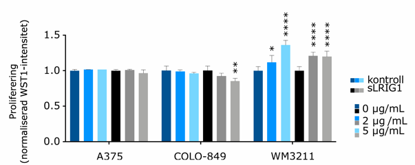
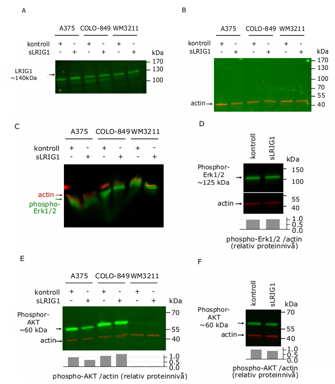
LRIG1 Expression and Functional Effects in Melanoma Subtypes
Triple wild-type melanoma lacks MAPK/ERK pathway mutations and effective treatments, while LRIG1 regulates EGFR upstream of MAPK/ERK and PI3K/AKT pathways. Hadi et al. investigated whether recombinant LRIG1 inhibits proliferation in BRAF-mutated (A375) and triple wild-type (colo-489, wm-3211) melanoma cells.
LRIG1 mRNA expression was significantly higher in metastatic triple wild-type melanoma compared to BRAF-mutated, RAS-mutated, and NF1-mutated subtypes. Recombinant LRIG1 dose-dependently inhibited proliferation in colo-849 cells but not in A375 or wm-3211 cells, with wm-3211 even showing stimulation (Fig. 1). Western blot analysis revealed low endogenous LRIG1 expression across all lines, undetectable phospho-EGFR, and failed/inconclusive phospho-ERK detection; notably, LRIG1 treatment suppressed phospho-AKT in A375 cells but not in colo-849 or wm-3211 cells (Fig. 2A-F).
Ask a Question
Write your own review
- You May Also Need

Description: Established from the primary tumor (right cervical) of a 64-year-old woman with cutaneous melanoma

Description: Established from the lymph node metastasis of a malignant melanoma from a 42-year-old Caucasian woman
Description: Species: human - male, 31 years old, CaucasianIsoenzyme: G6PD, BProduction: melaninHistopathology: melanoma
Description: Epstein-Barr virus-positive cell line established from peripheral blood in 1977 from male patient with melanoma

Description: Established from the primary tumor of a 58-year-old woman with melanoma in 1977

Description: Established from the primary (achromic) cutaneous tumor (left thigh) of a 26-year-old man with malignant melanoma (primary tumor histology: SSM level IV); same patient as cell line IGR-37; described ...
- Adipose Tissue-Derived Stem Cells
- Human Neurons
- Mouse Probe
- Whole Chromosome Painting Probes
- Hepatic Cells
- Renal Cells
- In Vitro ADME Kits
- Tissue Microarray
- Tissue Blocks
- Tissue Sections
- FFPE Cell Pellet
- Probe
- Centromere Probes
- Telomere Probes
- Satellite Enumeration Probes
- Subtelomere Specific Probes
- Bacterial Probes
- ISH/FISH Probes
- Exosome Isolation Kit
- Human Adult Stem Cells
- Mouse Stem Cells
- iPSCs
- Mouse Embryonic Stem Cells
- iPSC Differentiation Kits
- Mesenchymal Stem Cells
- Immortalized Human Cells
- Immortalized Murine Cells
- Cell Immortalization Kit
- Adipose Cells
- Cardiac Cells
- Dermal Cells
- Epidermal Cells
- Peripheral Blood Mononuclear Cells
- Umbilical Cord Cells
- Monkey Primary Cells
- Mouse Primary Cells
- Breast Tumor Cells
- Colorectal Tumor Cells
- Esophageal Tumor Cells
- Lung Tumor Cells
- Leukemia/Lymphoma/Myeloma Cells
- Ovarian Tumor Cells
- Pancreatic Tumor Cells
- Mouse Tumor Cells