
IGR-37
Cat.No.: CSC-C0343
Species: Homo sapiens (Human)
Source: Lymph Node Metastasis
Morphology: epithelial-like cells growing as monolayers
Culture Properties: monolayer
- Specification
- Background
- Scientific Data
- Q & A
- Customer Review
Immunology: cytokeratin -, cytokeratin-7 -, cytokeratin-8 -, cytokeratin-17 -, cytokeratin-18 -, cytokeratin-19
IGR-37 is a human melanoma cell line established from a lymph node metastasis (groin) of a 26‑year‑old male patient with malignant melanoma, primary tumor histology superficial spreading melanoma (SSM) level IV. The same patient also gave rise to the primary melanoma cell line IGR‑39 (CSC-C0344), making IGR‑37 a syngeneic, patient‑matched metastatic counterpart.
The primary IGR‑39 and metastatic IGR‑37 represent a unique, genetically correlated pair of human melanoma cells from the same donor. This allows direct molecular, phenotypic, and functional comparisons without confounding inter‑individual genetic variation. IGR‑37 consistently exhibits greater tumorigenicity in nude mice, forms larger tumors, and may harbor metastatic‑specific modifications such as altered ganglioside signatures (e.g., O‑acetylation of GM2, GD3 and GD2). This paired system is therefore an invaluable resource for identifying biomarkers of metastasis and for modeling the melanoma progression cascade.
IGR‑37 cells display an epithelial‑like morphology, a doubling time of approximately 48-72 hours, and a hypertriploid karyotype. They carry the BRAF V600E mutation but are wildtype for NRAS Q61, reflecting a common driver mutation in cutaneous melanoma. The line is inherently resistant to TRAIL‑induced apoptosis, an established model for studying apoptosis evasion and for testing sensitizing strategies.
Comparison of The Oncolytic Activity of A Replication-Competent and A Replication-Deficient Herpes Simplex Virus 1
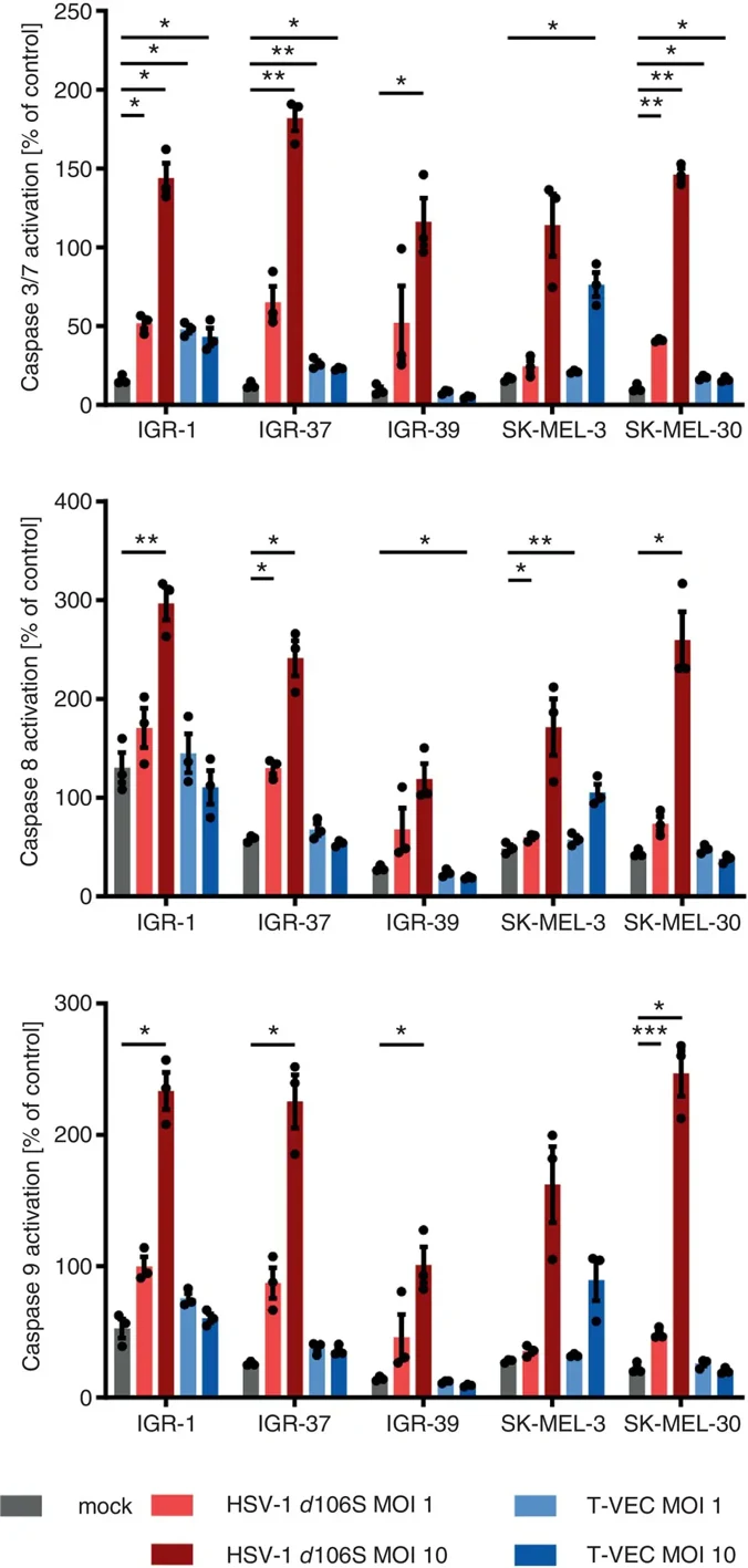
In 2015, the oncolytic herpes simplex virus 1 (HSV-1) T-VEC (talimogene laherparepvec) was approved for intratumoral injection in non-resectable malignant melanoma. To determine whether viral replication is required for oncolytic activity, we compared replication-deficient HSV-1 d106S with replication-competent T-VEC.
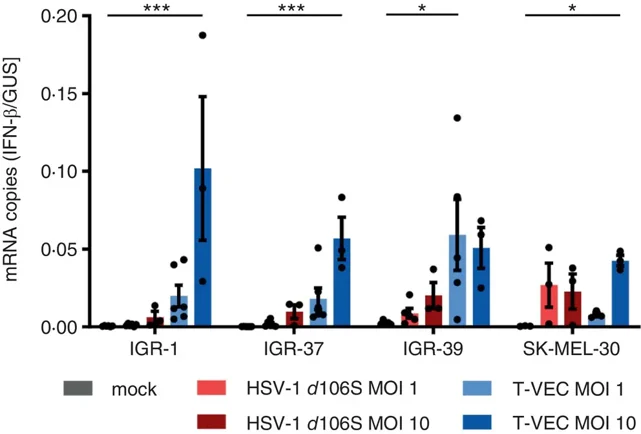
High infectious doses of HSV-1 d106S killed melanoma (n = 10), head-and-neck squamous cell carcinoma (n = 11), and chondrosarcoma cell lines (n = 2) significantly faster than T-VEC as measured by MTT metabolic activity, while low doses of T-VEC were more effective over time. HSV-1 d106S and, to a lesser extent T-VEC, triggered caspase-dependent early apoptosis as shown by pan-caspase inhibition and specific induction of caspases 3/7, 8, and 9. HSV-1 d106S induced a higher ratio of apoptosis-inducing infected cell protein (ICP) 0 to apoptosis-blocking ICP6 than T-VEC. T-VEC was oncolytic for an extended period of time as viral replication continued, which could be partially blocked by the antiviral drug aciclovir. High doses of T-VEC, but not HSV-1 d106S, increased interferon-β mRNA as part of the intrinsic immune response. When markers of immunogenic cell death were assessed, ATP was released more efficiently in the context of T-VEC than HSV-1 d106S infection, whereas HMGB1 was induced comparatively well.
Overall, the early oncolytic effect on three different tumor entities was stronger with the non-replicative strain, while the replication-competent virus elicited a stronger innate immune response and more pronounced immunogenic cell death.
Ask a Question
Write your own review
- You May Also Need

Description: Established in 1990 from the right axillary lymph node of a 48-year-old woman with metastatic malignant melanoma in the right chest

Description: Established from the primary tumor (right cervical) of a 64-year-old woman with cutaneous melanoma


Description: Established from the lymph node metastasis of a malignant melanoma from a 42-year-old Caucasian woman

- Adipose Tissue-Derived Stem Cells
- Human Neurons
- Mouse Probe
- Whole Chromosome Painting Probes
- Hepatic Cells
- Renal Cells
- In Vitro ADME Kits
- Tissue Microarray
- Tissue Blocks
- Tissue Sections
- FFPE Cell Pellet
- Probe
- Centromere Probes
- Telomere Probes
- Satellite Enumeration Probes
- Subtelomere Specific Probes
- Bacterial Probes
- ISH/FISH Probes
- Exosome Isolation Kit
- Human Adult Stem Cells
- Mouse Stem Cells
- iPSCs
- Mouse Embryonic Stem Cells
- iPSC Differentiation Kits
- Mesenchymal Stem Cells
- Immortalized Human Cells
- Immortalized Murine Cells
- Cell Immortalization Kit
- Adipose Cells
- Cardiac Cells
- Dermal Cells
- Epidermal Cells
- Peripheral Blood Mononuclear Cells
- Umbilical Cord Cells
- Monkey Primary Cells
- Mouse Primary Cells
- Breast Tumor Cells
- Colorectal Tumor Cells
- Esophageal Tumor Cells
- Lung Tumor Cells
- Leukemia/Lymphoma/Myeloma Cells
- Ovarian Tumor Cells
- Pancreatic Tumor Cells
- Mouse Tumor Cells