A2058
Cat.No.: CSC-C6976J
Species: Homo sapiens (Human)
Source: Lymph Node Metastasis
Morphology: Epithelial-Like
- Specification
- Background
- Scientific Data
- Q & A
- Customer Review
A2058 is a human malignant melanoma cell line derived from the metastatic site of an individual with cutaneous melanoma. The cell line models advanced melanoma and is often utilized as an in vitro system for studying the molecular processes associated with melanoma progression, invasion, and therapeutic resistance. Morphologically, A2058 cells are adherent monolayers with an epithelial-like to spindle-shaped appearance, and they exhibit strong proliferation in standard culture conditions, commonly using DMEM with fetal bovine serum as a supplement.
On the molecular level, A2058 harbors activating mutations in key oncogenic signaling pathways, such as the MAPK pathway, which is commonly dysregulated in melanoma. Studies show that this cell line exhibits modified expression of melanoma markers and its signaling components related to the aggressive phenotype and invasive potential making it ideal for research on pathway-specific inhibitors and resistance mechanisms. Functionally, A2058 cells exhibit robust migratory and invasive properties and can readily form tumors in xenograft models, reflecting its metastatic origin. The cell line is commonly used in studies of melanoma cell motility, epithelial-mesenchymal transition, and interactions with the tumor microenvironment.
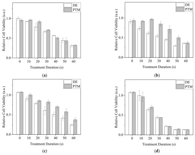
Viability of A2058 Cancer Cells with Direct and Indirect Ar-CAP Treatments
CAP generates RONS and is a potential cancer therapy tool. Here, Chen's team measured reactive species intensity in plasma gas and RONS concentrations in PBS and cell culture medium. They assessed cell viability after treating cells with plasma in PBS and medium under various conditions and compared the sensitivities of different cells.
A2058 cells were treated with Ar-CAP using two methods: direct exposure (DE) and plasma-treated medium (PTM). For DE, cells were exposed to Ar-CAP in medium volumes of 30, 50, 100, and 150 µL for 0 to 60 seconds (DE0 to DE60) and cultured for 20 hours. For PTM, medium volumes of 30, 50, 100, and 150 µL were treated with plasma for 0 to 60 seconds (P0 to P60) before culturing cells for 20 hours. Cell viability was measured by MTT assay (Fig. 1). In 30 and 150 µL, both DE and PTM decreased cell viability with longer treatment times, but effects were similar. In 50 and 100 µL, DE had stronger lethal effects than PTM. The 150 µL treatments had the most severe lethal effects, likely due to higher plasma intensity generating more RONS and causing stronger oxidative stress.
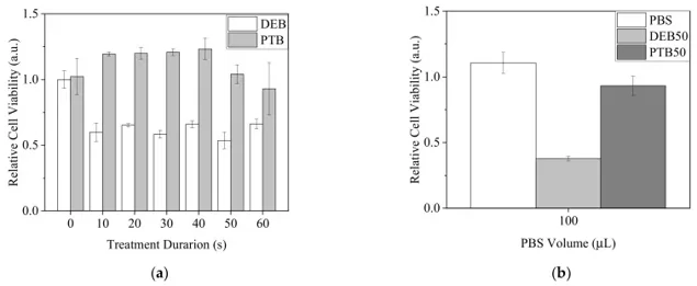
To investigate short-lived RONS, cells were treated with direct plasma exposure in PBS (DEB) for 0 to 60 seconds or with plasma-treated PBS (PTB) at 30 µL. After 20 minutes, treated PBS was replaced with complete medium and cells were cultured for 20 hours. MTT assays showed that DEB significantly reduced cell viability, with no clear changes over different treatment durations. PTB had minor effects or increased cell viability (Fig. 2a). When 100 µL was used for 50 seconds of plasma exposure, DEB showed a higher lethal effect than PTB (Fig. 2b). These results suggest that short-lived RONS strongly affect cells with DEB treatment.
Ask a Question
Write your own review
- You May Also Need

Description: Established from the primary tumor (right cervical) of a 64-year-old woman with cutaneous melanoma

Description: Established from the lymph node metastasis of a malignant melanoma from a 42-year-old Caucasian woman
Description: Species: human - male, 31 years old, CaucasianIsoenzyme: G6PD, BProduction: melaninHistopathology: melanoma

Description: Epstein-Barr virus-positive cell line established from peripheral blood in 1977 from male patient with melanoma

Description: Established from the primary tumor of a 58-year-old woman with melanoma in 1977

Description: Established from the primary (achromic) cutaneous tumor (left thigh) of a 26-year-old man with malignant melanoma (primary tumor histology: SSM level IV); same patient as cell line IGR-37; described ...
- Adipose Tissue-Derived Stem Cells
- Human Neurons
- Mouse Probe
- Whole Chromosome Painting Probes
- Hepatic Cells
- Renal Cells
- In Vitro ADME Kits
- Tissue Microarray
- Tissue Blocks
- Tissue Sections
- FFPE Cell Pellet
- Probe
- Centromere Probes
- Telomere Probes
- Satellite Enumeration Probes
- Subtelomere Specific Probes
- Bacterial Probes
- ISH/FISH Probes
- Exosome Isolation Kit
- Human Adult Stem Cells
- Mouse Stem Cells
- iPSCs
- Mouse Embryonic Stem Cells
- iPSC Differentiation Kits
- Mesenchymal Stem Cells
- Immortalized Human Cells
- Immortalized Murine Cells
- Cell Immortalization Kit
- Adipose Cells
- Cardiac Cells
- Dermal Cells
- Epidermal Cells
- Peripheral Blood Mononuclear Cells
- Umbilical Cord Cells
- Monkey Primary Cells
- Mouse Primary Cells
- Breast Tumor Cells
- Colorectal Tumor Cells
- Esophageal Tumor Cells
- Lung Tumor Cells
- Leukemia/Lymphoma/Myeloma Cells
- Ovarian Tumor Cells
- Pancreatic Tumor Cells
- Mouse Tumor Cells