GS-HepG2
Cat.No.: CSC-C6373J
Species: Homo sapiens (Human)
Source: Liver
Morphology: epithelial-like
Culture Properties: Adherent cells
- Specification
- Background
- Scientific Data
- Q & A
- Customer Review
Store in liquid nitrogen.
GS-HepG2 is a glutamine synthetase-expressing mutant cell line derived from HepG2. HepG2 itself is a commonly employed cell line derived from human hepatocellular carcinoma (liver tumor) that displays many hepatocyte-like characteristics, such as albumin secretion and expression of drug metabolizing enzymes. GS-HepG2 cells were generated via stable transfection with glutamine synthetase (GS, also known as GLUL), which allows for easier metabolism of ammonia and selection in glutamine-free media. Parental HepG2 cells lack the ability to detoxify ammonia.
They metabolize nitrogenous waste more efficiently than their parent cell line, and have been used as part of bioartificial liver systems and assay platforms for hepatocyte function. GS-HepG2 cells have been shown to lower ammonia concentrations in bioreactors and support sustained cultures. Engineered derivatives of GS-HepG2 such as GS-3A4-HepG2 are also used in research settings as a model for drug metabolism and cytochrome P450 induction.
Promoter Methylation and H3K27 Deacetylation Co-Regulate VIPR1 Transcription in Hepatocellular Carcinoma
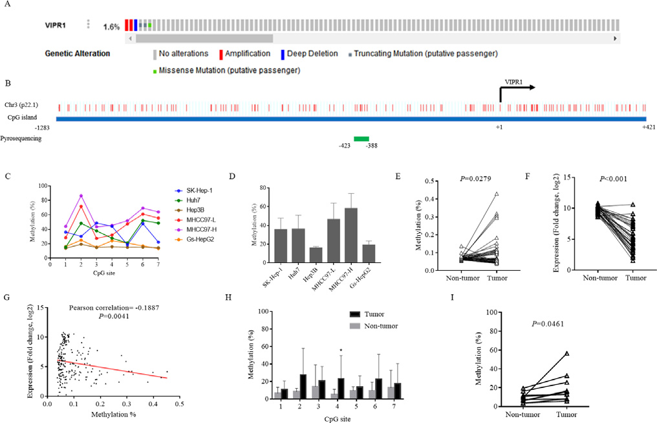
Vasoactive intestinal peptide receptor 1 (VIPR1) is differentially expressed in human cancers. To uncover its clinical relevance and mechanism of transcriptional regulation in hepatocellular carcinoma (HCC), Lu et al. first identified VIPR1 CpG island by UCSC Genome Browser.
After selection, methylation status of seven CpG sites was examined by pyrosequencing (Fig. 1B). DNA methylation was detected at seven CpG sites in all of six HCC cell lines (SK-Hep-1, Huh7, Hep3B, MHCC97-L, MHCC97-H, Gs-HepG2) (Fig. 1C). Among these six cell lines, methylation levels of SK-Hep-1, Huh7, MHCC97-L and MHCC97-H cells were higher than those of Hep3B and Gs-HepG2 cells (Fig. 1D). Methylation levels of VIPR1 in 41 pairs of HCC and paracancerous tissues from MethHC database were significantly higher in HCC tissues than those in corresponding non-cancerous tissues (Fig. 1E), while mRNA levels were significantly lower in HCC than those in non-cancerous tissues (Fig. 1F). Further correlation analysis was performed between methylation and mRNA expression levels in 41 paired adjacent normal tissues and HCC tissues, as well as 189 HCC tissues from MethHC database. A negative correlation between methylation and mRNA levels of VIPR1 was observed in these tissues (Fig. 1G). Another 10 pairs of matched HCC tissues and paracancerous tissues were recruited to validate the methylation status of VIPR1. As shown in Figure 1H, high methylation levels were detected at each CpG locus in HCC tissues, while low methylation levels were observed in non-cancerous tissues. The mean levels of methylation at seven CpG sites in HCC tissues were higher than those in corresponding non-cancerous tissues (Fig. 1I), which was consistent with bioinformatics analyses.
Ask a Question
Write your own review
- You May Also Need
Description: Ito (fat-storing) cells, stellate-shaped mesenchymal cells that exist in the space of Disse of the liver and contain many fat droplets in cytoplasm.

Description: This is one cell line out of a series of glioblastoma cell lines established by PD Dr. Michael Linnebacher.
Description: Human bile duct cell line established from ascites of the tumor patient who had differentiated adenocarcinoma.


Description: Human cell line derived from cholangiocellular carcinoma. Cell growth is slow.
- Adipose Tissue-Derived Stem Cells
- Human Neurons
- Mouse Probe
- Whole Chromosome Painting Probes
- Hepatic Cells
- Renal Cells
- In Vitro ADME Kits
- Tissue Microarray
- Tissue Blocks
- Tissue Sections
- FFPE Cell Pellet
- Probe
- Centromere Probes
- Telomere Probes
- Satellite Enumeration Probes
- Subtelomere Specific Probes
- Bacterial Probes
- ISH/FISH Probes
- Exosome Isolation Kit
- Human Adult Stem Cells
- Mouse Stem Cells
- iPSCs
- Mouse Embryonic Stem Cells
- iPSC Differentiation Kits
- Mesenchymal Stem Cells
- Immortalized Human Cells
- Immortalized Murine Cells
- Cell Immortalization Kit
- Adipose Cells
- Cardiac Cells
- Dermal Cells
- Epidermal Cells
- Peripheral Blood Mononuclear Cells
- Umbilical Cord Cells
- Monkey Primary Cells
- Mouse Primary Cells
- Breast Tumor Cells
- Colorectal Tumor Cells
- Esophageal Tumor Cells
- Lung Tumor Cells
- Leukemia/Lymphoma/Myeloma Cells
- Ovarian Tumor Cells
- Pancreatic Tumor Cells
- Mouse Tumor Cells