
YNH-1
Cat.No.: CSC-C0673
Species: Homo sapiens (Human)
Source: Blood; Peripheral Blood
Morphology: single round cells growing in suspension, some cells are loosely adherent
Culture Properties: suspension
- Specification
- Background
- Scientific Data
- Q & A
- Customer Review
Immunology: CD3 -, CD4 +, CD13 +, CD14 -, CD15 -, CD19 -, CD20 -, CD33 +, CD34 +, cyCD68 +, HLA-DR -
Viruses: PCR: EBV -, HBV -, HCV -, HIV -, HTLV-I/-II -
YNH-1 is a human acute myeloid leukemia (AML) cell line. This cell line was derived from peripheral blood of a 46-year-old male patient with acute myeloid leukemia (AML, FAB M1 type). It was originally created as a tool to study leukemia with rare chromosomal translocations, specifically it had t(16;21)(p11;q22). YNH-1 cells proliferate as immature cells of myeloblastoid lineage in cell culture and display a doubling time of approximately 80-82 hours. In order to maintain proliferation these cells require exogenous cytokines such as granulocyte colony-stimulating factor (G-CSF), granulocyte-macrophage colony-stimulating factor (GM-CSF) or interleukin-3 (IL-3). YNH-1 cells display typical immunophenotype markers of myeloid cells such as CD13, CD33 and CD34. Positive myeloperoxidase activity was detected.
The t(16;21) translocation in YNH-1 cells results in the fusion of the FUS and ERG genes, creating the FUS-ERG (TLS-ERG) fusion. FUS-ERG is a known oncogene that contributes to leukemogenesis and poor prognosis. These cells have been used extensively to study rare subtypes of AML. YNH-1 has been used as a tool to study many facets of leukemia including; chromosomal translocation, oncogenic properties of translocation products, cytokine-dependent proliferation of leukemia cells, and signaling in AML.
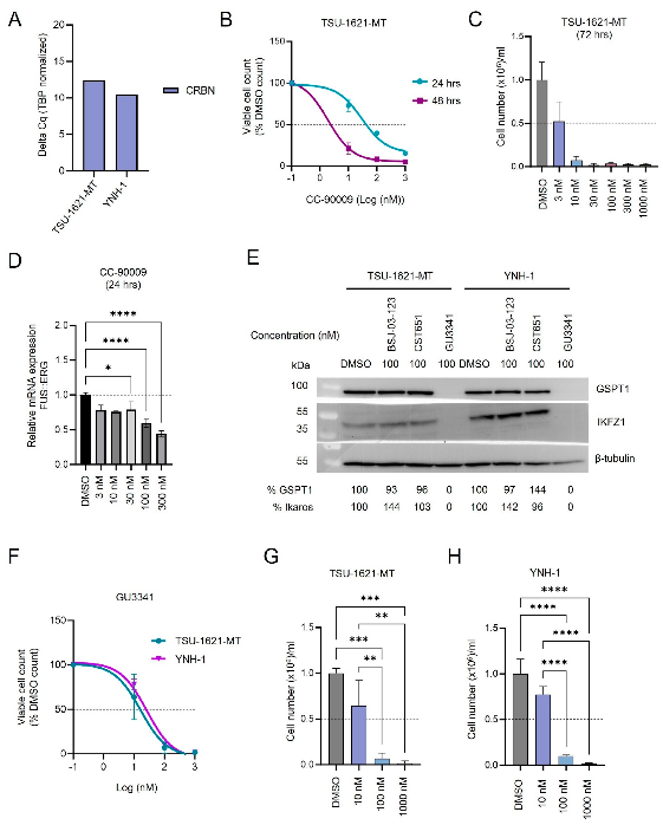
CC-90009 and GU3341 PROTACs Induce Anti-AML Activity in FUS::ERG Cell Lines
PROTACs selectively degrade target proteins via the ubiquitin-proteasome system, showing efficacy in adult AML but remaining unexplored in pediatric AML. Perzolli et al. demonstrates potent anti-AML effects through GSPT1 degradation by CC-90009 or off-target GU3341 activity.
Given CC-90009-induced ERG degradation, they examined its impact on FUSERG-positive t(16;21)(p11;q22) AML, a subtype with particularly poor prognosis. Both FUSERG cell lines (TSU-1621-MT, YNH-1) expressed significant CRBN levels (Fig. 1A). CC-90009 showed stronger antiproliferative activity in TSU-1621-MT versus Kasumi-1 cells (ED50: 30.2 ± 13 nM at 24 h, 2.0 ± 0.8 nM at 48 h), with potent cytotoxicity at 10 nM by 72 h (Fig. 1B and C). Notably, FUS::ERG fusion transcript levels significantly decreased after 24 h CC-90009 treatment (Fig. 1D).
Immunoblotting confirmed GU3341-induced GSPT1 degradation in both t(16;21) lines (Fig. 1E). GU3341 exhibited strong cytotoxicity, achieving >90% killing at 100 nM and nearly 100% at 1000 nM by 72 h in both lines (Fig. 1F-H). Both FUSERG lines were 5-10-fold more sensitive to GU3341 than RUNX1RUNX1T1 lines (ED50: Kasumi-1 = 164 ± 2 nM; TSU-1621-MT = 16 ± 2 nM; YNH-1 = 29 ± 2 nM). These findings indicate that t(16;21)(p11;q22) AML is particularly sensitive to GSPT1 degraders.
Ask a Question
Write your own review
- You May Also Need

Description: Established in 2007 from the bone marrow mononuclear cells of an 82-year-old Japanese man with diffuse large B-cell lymphoma in the leukemic phase

Description: Established from the bone marrow of a 28-year-old man who developed the terminal leukemic phase of lymphosarcoma in 1976

Description: This cell line was derived from the bone marrow aspirate of a 59 year old male with erythroleukemia that became acute myelogenous leukaemia.The cells form colonies in soft-agar in the presence of ...

Description: Established from the pleural effusion of a 24-year-old woman with recurrent anaplastic large cell lymphoma (ALCL); cells were described to clonally derive from T-lineage lymphoid cells and to be ...

Description: Established from a 37-year-old man at second (refractory/terminal) relapse of Hodgkin lymphoma (nodular sclerosing -> lymphocyte depleted/stage IIISA -> stage IV) after both combined chemo- and ...

Description: Established from the peripheral blood of a 10-year-old Caucasian boy with acute lymphoblastic leukemia (pre B-ALL) at diagnosis in 1993
- Adipose Tissue-Derived Stem Cells
- Human Neurons
- Mouse Probe
- Whole Chromosome Painting Probes
- Hepatic Cells
- Renal Cells
- In Vitro ADME Kits
- Tissue Microarray
- Tissue Blocks
- Tissue Sections
- FFPE Cell Pellet
- Probe
- Centromere Probes
- Telomere Probes
- Satellite Enumeration Probes
- Subtelomere Specific Probes
- Bacterial Probes
- ISH/FISH Probes
- Exosome Isolation Kit
- Human Adult Stem Cells
- Mouse Stem Cells
- iPSCs
- Mouse Embryonic Stem Cells
- iPSC Differentiation Kits
- Mesenchymal Stem Cells
- Immortalized Human Cells
- Immortalized Murine Cells
- Cell Immortalization Kit
- Adipose Cells
- Cardiac Cells
- Dermal Cells
- Epidermal Cells
- Peripheral Blood Mononuclear Cells
- Umbilical Cord Cells
- Monkey Primary Cells
- Mouse Primary Cells
- Breast Tumor Cells
- Colorectal Tumor Cells
- Esophageal Tumor Cells
- Lung Tumor Cells
- Leukemia/Lymphoma/Myeloma Cells
- Ovarian Tumor Cells
- Pancreatic Tumor Cells
- Mouse Tumor Cells