
TOM-1
Cat.No.: CSC-C0583
Species: Homo sapiens (Human)
Source: Bone Marrow
Morphology: small round cells growing in suspension singly or in clumps, some cells are loosely adherent
Culture Properties: suspension
- Specification
- Background
- Scientific Data
- Q & A
- Customer Review
Immunology: CD2 -, CD3 -, CD4 -, CD7 -, CD10 +, CD13 +, CD19 +, CD20 +, CD34 +, CD37 +, CD38 +, cyCD79a +, CD80 -, CD138 -, HLA-DR +, sm/cyIgG -, sm/cyIgM -, sm/cykappa -, sm/cylambda -
Viruses:
The TOM-1 cell line is a human B-cell precursor leukemia line established in 1983 from the bone marrow of a 54-year-old female patient with refractory Philadelphia chromosome-positive acute lymphoblastic leukemia (Ph+ ALL). It is characterized by a hyperdiploid karyotype with a t(9;22) translocation, resulting in the expression of the p190 BCR-ABL1 fusion protein (e1a2 junction). Morphologically, TOM-1 cells are small, round lymphocytes that grow in suspension, either singly or in loose clusters, though some may show loose adherence. They are positive for HLA-DR, CD10, CD19, CD20, CD34, and terminal deoxynucleotidyl transferase (TdT), but negative for surface and cytoplasmic immunoglobulins, confirming their early pre-B cell stage. Culturally, they are maintained in a nutrient-rich basal medium supplemented with serum under standard conditions (37°C, 5% CO₂). The line has a doubling time of approximately 50-70 hours and is sub-cultured upon reaching appropriate density.
TOM-1 is a valuable model for studying Ph+ ALL pathogenesis, BCR-ABL1 signaling, tyrosine kinase inhibitor sensitivity, and mechanisms of leukemic stem cell differentiation and drug resistance.
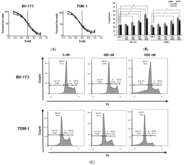
Biological Effects of LIMKi in BCR::ABL+ Cell Lines
Despite advances with TKIs, Philadelphia chromosome-positive B-cell acute lymphoblastic leukemia (Ph+ B-ALL) remains a high-risk malignancy, necessitating less toxic strategies, particularly for elderly patients. Berrou et al. evaluated targeting LIMK1/2-key downstream effectors of BCR::ABL that regulate apoptosis via cofilin phosphorylation.
As BCR::ABL activates the ROCK-LIMK axis, they investigated the efficacy of the LIMK inhibitor CEL_Amide in Ph+ B-ALL cell lines (TOM-1, BV-173). CEL_Amide significantly inhibited cell viability with IC50s of 580 nM (TOM-1) and 1090 nM (BV-173) (Fig. 1A). Treatment induced dose-dependent apoptosis, reaching 45% (TOM-1) and 50% (BV-173) at 48 h (Fig. 1B), and caused G1/S cell cycle arrest (Fig. 1C). These findings suggest that LIMK inhibition disrupts critical survival pathways in Ph+ B-ALL.
Ask a Question
Write your own review
- You May Also Need

Description: Established in 2007 from the bone marrow mononuclear cells of an 82-year-old Japanese man with diffuse large B-cell lymphoma in the leukemic phase

Description: Established from the bone marrow of a 28-year-old man who developed the terminal leukemic phase of lymphosarcoma in 1976

Description: This cell line was derived from the bone marrow aspirate of a 59 year old male with erythroleukemia that became acute myelogenous leukaemia.The cells form colonies in soft-agar in the presence of ...

Description: Established from the pleural effusion of a 24-year-old woman with recurrent anaplastic large cell lymphoma (ALCL); cells were described to clonally derive from T-lineage lymphoid cells and to be ...

Description: Established from a 37-year-old man at second (refractory/terminal) relapse of Hodgkin lymphoma (nodular sclerosing -> lymphocyte depleted/stage IIISA -> stage IV) after both combined chemo- and ...

Description: Established from the peripheral blood of a 10-year-old Caucasian boy with acute lymphoblastic leukemia (pre B-ALL) at diagnosis in 1993
- Adipose Tissue-Derived Stem Cells
- Human Neurons
- Mouse Probe
- Whole Chromosome Painting Probes
- Hepatic Cells
- Renal Cells
- In Vitro ADME Kits
- Tissue Microarray
- Tissue Blocks
- Tissue Sections
- FFPE Cell Pellet
- Probe
- Centromere Probes
- Telomere Probes
- Satellite Enumeration Probes
- Subtelomere Specific Probes
- Bacterial Probes
- ISH/FISH Probes
- Exosome Isolation Kit
- Human Adult Stem Cells
- Mouse Stem Cells
- iPSCs
- Mouse Embryonic Stem Cells
- iPSC Differentiation Kits
- Mesenchymal Stem Cells
- Immortalized Human Cells
- Immortalized Murine Cells
- Cell Immortalization Kit
- Adipose Cells
- Cardiac Cells
- Dermal Cells
- Epidermal Cells
- Peripheral Blood Mononuclear Cells
- Umbilical Cord Cells
- Monkey Primary Cells
- Mouse Primary Cells
- Breast Tumor Cells
- Colorectal Tumor Cells
- Esophageal Tumor Cells
- Lung Tumor Cells
- Leukemia/Lymphoma/Myeloma Cells
- Ovarian Tumor Cells
- Pancreatic Tumor Cells
- Mouse Tumor Cells