KMOE-2
Cat.No.: CSC-C0213
Species: Homo sapiens (Human)
Source: Blood; Peripheral Blood
Morphology: single, large, round to oval cells in suspension
- Specification
- Background
- Scientific Data
- Q & A
- Customer Review
KMOE-2 is a continuous human erythroid leukaemia cell line established in 1978 from peripheral blood of a 2-year-old female patient with acute erythremia (Di Guglielmo's illness). It has a hypotetraploid karyotype (modal number 82, range 80-88) and develops in suspension as solitary, big, round to oval cells.
KMOE-2 cells are cultured in conventional conditions (37°C, 5% CO2) in a nutrient-rich basal medium supplemented with serum. Their doubling time is about 40-50 h. They are seeded at a density of 0.2-0.4 × 10 6 cells/mL and achieve a peak density of about 0.6 × 10 6 cells/mL.
One of the characteristics of KMOE-2 is its erythroid differentiation potential. When treated with metabolic inhibitors such as cytosine arabinoside, the cells could synthesize hemoglobin and show benzidine-positive phenotypes. Therefore, this cell line is a useful model for researching erythropoiesis, processes of leukemic cell differentiation and screening of new therapeutic drugs against hematologic malignancies.
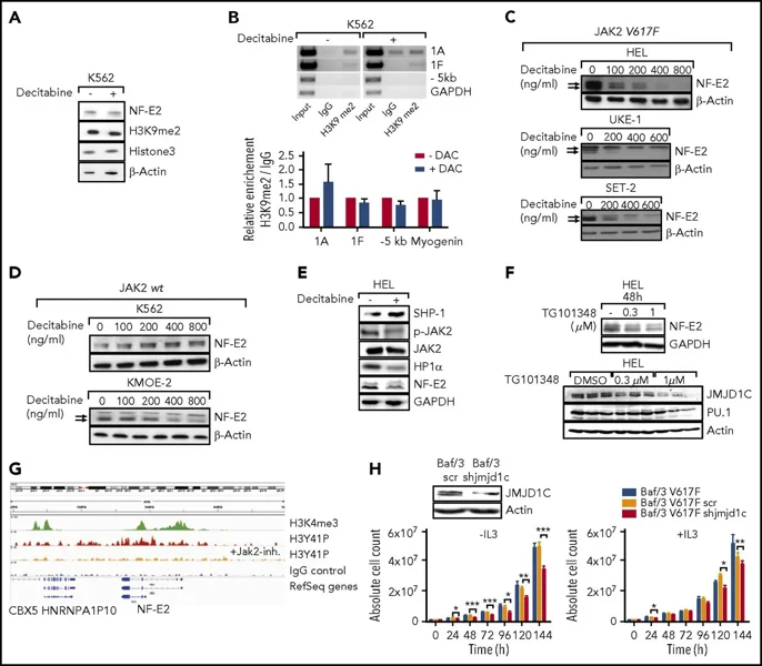
NFE2 Is Regulated via JAK2-Mediated Histone Tyrosine Phosphorylation
NFE2 is frequently overexpressed in myeloproliferative neoplasms (MPNs) and drives disease progression in murine models, yet its upstream regulators and downstream effectors remain unclear. Peeken et al. investigated how epigenetic modifications, specifically histone methylation and phosphorylation, regulate NFE2 expression.
They examined the role of JAK2-mediated phosphorylation. JAK2 phosphorylates Histone H3 at Tyrosine 41 (H3Y41), disrupting Heterochromatin Protein 1α (HP1α) binding. The NFE2-1A promoter harbored phospho-H3Y41, and this mark was significantly reduced by decitabine treatment.
To assess the role of JAK2 mutational status, they compared JAK2V617F mutant cell lines (HEL, UKE-1, SET-2) with JAK2 wild-type (wt) lines (K562, KMOE-2). Decitabine effectively silenced NFE2 and reduced H3K9me2 at its promoters in JAK2V617F mutant cells (Fig. 1A-C). In contrast, decitabine had no significant effect on NFE2 levels or H3K9me2 marks in JAK2wt cells (Fig. 1A, B, D). These findings indicate that decitabine selectively targets NFE2 expression in cells harboring the JAK2V617F mutation.
Ask a Question
Write your own review
- You May Also Need

Description: Established in 2007 from the bone marrow mononuclear cells of an 82-year-old Japanese man with diffuse large B-cell lymphoma in the leukemic phase

Description: Established from the bone marrow of a 28-year-old man who developed the terminal leukemic phase of lymphosarcoma in 1976

Description: This cell line was derived from the bone marrow aspirate of a 59 year old male with erythroleukemia that became acute myelogenous leukaemia.The cells form colonies in soft-agar in the presence of ...

Description: Established from the pleural effusion of a 24-year-old woman with recurrent anaplastic large cell lymphoma (ALCL); cells were described to clonally derive from T-lineage lymphoid cells and to be ...

Description: Established from a 37-year-old man at second (refractory/terminal) relapse of Hodgkin lymphoma (nodular sclerosing -> lymphocyte depleted/stage IIISA -> stage IV) after both combined chemo- and ...

Description: Established from the peripheral blood of a 10-year-old Caucasian boy with acute lymphoblastic leukemia (pre B-ALL) at diagnosis in 1993
- Adipose Tissue-Derived Stem Cells
- Human Neurons
- Mouse Probe
- Whole Chromosome Painting Probes
- Hepatic Cells
- Renal Cells
- In Vitro ADME Kits
- Tissue Microarray
- Tissue Blocks
- Tissue Sections
- FFPE Cell Pellet
- Probe
- Centromere Probes
- Telomere Probes
- Satellite Enumeration Probes
- Subtelomere Specific Probes
- Bacterial Probes
- ISH/FISH Probes
- Exosome Isolation Kit
- Human Adult Stem Cells
- Mouse Stem Cells
- iPSCs
- Mouse Embryonic Stem Cells
- iPSC Differentiation Kits
- Mesenchymal Stem Cells
- Immortalized Human Cells
- Immortalized Murine Cells
- Cell Immortalization Kit
- Adipose Cells
- Cardiac Cells
- Dermal Cells
- Epidermal Cells
- Peripheral Blood Mononuclear Cells
- Umbilical Cord Cells
- Monkey Primary Cells
- Mouse Primary Cells
- Breast Tumor Cells
- Colorectal Tumor Cells
- Esophageal Tumor Cells
- Lung Tumor Cells
- Leukemia/Lymphoma/Myeloma Cells
- Ovarian Tumor Cells
- Pancreatic Tumor Cells
- Mouse Tumor Cells