
- Specification
- Background
- Scientific Data
- Q & A
- Customer Review
MOG-G-CCM is a mouse glioma cell line developed from genetically altered animal models of gliomagenesis and is often used as an in vitro model for aggressive brain cancers, especially glioblastoma-like characteristics. This cell line has good proliferative capability and adherent growth properties and shows spindle-shaped or mixed morphology, which is compatible with glial tumor genesis.
MOG-G-CCM cells retain important molecular and immunological properties of CNS malignancies, and hence, provide a useful tool to study tumor biology in a syngeneic or immunocompetent setting. They are commonly utilized in studies of glioma cell proliferation, invasion, and tumor-microenvironment interactions such as crosstalk with microglia, astrocytes, and infiltrating immune cells.
Because of their murine origin, MOG-G-CCM cells are especially ideal for preclinical research involving immune-oncology and development of immunotherapy, including evaluation of checkpoint inhibitors, cytokine signaling pathways, and tumor-associated immune responses. They are also used in mechanistic studies of glioma growth, signaling system dysregulation and treatment resistance.
APR-246 Combined with Epigenetic Modulators Reduces Glioblastoma Clonogenicity
Glioblastoma remains refractory to conventional surgery, radiotherapy, and temozolomide chemotherapy, necessitating novel therapeutic strategies. Rosa et al. explored the combination of APR‑246, an agent aimed at restoring mutant p53 function, with epigenetic modulators DZ Nep (EZH2 inhibitor) and panobinostat, alongside temozolomide. Using the Chou-Talalay method, APR‑246 demonstrated additive interactions with these agents, reducing clonogenicity and inducing apoptosis irrespective of p53 status.
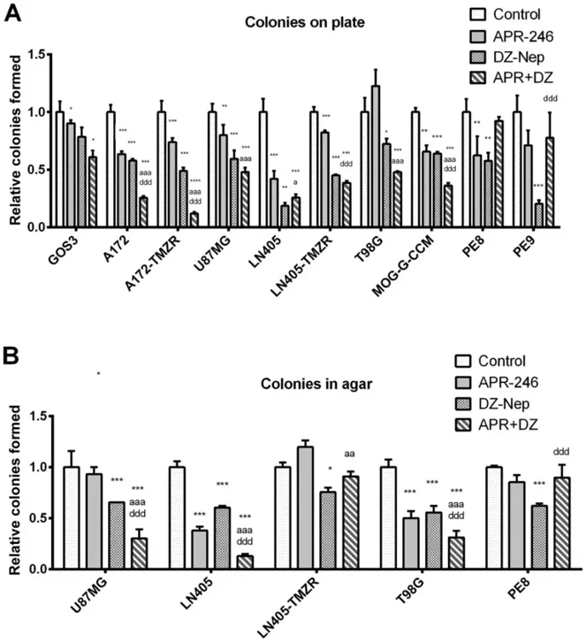
In adherent colony formation assays, GOS‑3 cells showed a significant reduction in colony-forming ability following combined DZ‑Nep and APR‑246 treatment compared to controls (Fig. 1A). PE8 and PE9 primary tumor cells exhibited no significant changes, whereas LN405 cells were more sensitive than their temozolomide-resistant counterpart (LN405‑TMZR) to single-agent or combined treatment. Similarly, A172‑TMZR cells were more responsive than parental A172 cells, forming fewer colonies under combination therapy. Additional cell lines (U87MG, T98G, MOG‑G‑CCM) also showed marked suppression of colony formation with dual treatment (Fig. 1A).
Soft agar assays confirmed these findings, with significant decreases in colony numbers for T98G, U87MG, and LN405 treated with APR‑246 plus DZ‑Nep (Fig. 1B). No significant reductions were observed in LN405‑TMZR or PE8 cells, and LN405‑TMZR showed increased colony formation upon APR‑246 monotherapy. These results highlight the potential of combining APR‑246 with epigenetic inhibitors to suppress glioblastoma growth, particularly in select resistant contexts.
Ask a Question
Write your own review
Description: Established from the temporal lobe brain biopsy from a 63-year-old man with glioblastoma multiforme in 1990 (prior to treatment)
Description: Established from the biopsy specimens from a 42-year-old woman with glioma (histologically defined as glioblastoma)
Description: Established from the frontal brain lobe of a 54-year-old woman with glioblastoma multiforme in 1989 (pre-treatment)
Description: Derived from an adult female with glioblastoma multiforme treated with local brain irradiation and multi drug chemotherapy. By flow cytometry these cells were almost 100% positive for MHC Class I.
- Adipose Tissue-Derived Stem Cells
- Human Neurons
- Mouse Probe
- Whole Chromosome Painting Probes
- Hepatic Cells
- Renal Cells
- In Vitro ADME Kits
- Tissue Microarray
- Tissue Blocks
- Tissue Sections
- FFPE Cell Pellet
- Probe
- Centromere Probes
- Telomere Probes
- Satellite Enumeration Probes
- Subtelomere Specific Probes
- Bacterial Probes
- ISH/FISH Probes
- Exosome Isolation Kit
- Human Adult Stem Cells
- Mouse Stem Cells
- iPSCs
- Mouse Embryonic Stem Cells
- iPSC Differentiation Kits
- Mesenchymal Stem Cells
- Immortalized Human Cells
- Immortalized Murine Cells
- Cell Immortalization Kit
- Adipose Cells
- Cardiac Cells
- Dermal Cells
- Epidermal Cells
- Peripheral Blood Mononuclear Cells
- Umbilical Cord Cells
- Monkey Primary Cells
- Mouse Primary Cells
- Breast Tumor Cells
- Colorectal Tumor Cells
- Esophageal Tumor Cells
- Lung Tumor Cells
- Leukemia/Lymphoma/Myeloma Cells
- Ovarian Tumor Cells
- Pancreatic Tumor Cells
- Mouse Tumor Cells