NBL-S
Cat.No.: CSC-C0638
Species: Homo sapiens (Human)
Morphology: polymorph cells growing adherently in monolayers
Culture Properties: monolayer
- Specification
- Background
- Scientific Data
- Q & A
- Customer Review
Immunology: cytokeratin -, cytokeratin-7 -, cytokeratin-8 -, cytokeratin-17 -, cytokeratin-18 -, desmin -, endothel -, EpCAM -, GFAP -, neurofila
The NBL-S cell line originated from the primary tumor of a neuroblastoma patient whose tumor developed from neural crest cells responsible for producing many embryonic tissues and organs including those of the nervous and endocrine systems. Neuroblastoma presents as a malignant tumor developing from the sympathetic nervous system which most frequently appears in pediatric patients and ranks as one of their most common cancers. Investigations revealed that this patient's tumor lacked MYCN gene amplification but displayed an extended MYCN protein half-life of about 100 minutes which exceeds the typical 30-minute half-life observed in NB cell lines. The exceptional genetic trait of NBL-S establishes it as a significant research instrument for studying neuroblastoma tumor biology. Metabolically, NBL-S cells demonstrate robust glycolytic activity which allows them to generate ATP when oxidative phosphorylation is blocked while simultaneously producing substantial lactic acid. NBL-S cells primarily utilize glycolysis for energy production which demonstrates their limited connection to the Warburg effect. The NBL-S cell line serves as a fundamental research tool focusing on neuroblastoma pathogenesis, drug testing, and therapeutic development in cancer biology studies.
HCP5 could Promote the Proliferation of Neuroblastoma Cells
Among pediatric patients, neuroblastoma represents the most frequently occurring solid tumor. Research indicated that long-chain noncoding RNA (lncRNA) HCP5 played a key role in tumor development yet its specific function in NB was not yet determined. Zhu's team plans to investigate HCP5's function in NB and identify its molecular mechanism.
HCP5 expression levels in neuroblastoma tissues exceeded those in paracancerous tissues as demonstrated by RT-PCR results (Fig. 1 A). SHSY-5Y and Kelly cells with elevated HCP5 expression demonstrated superior clone formation abilities compared to NBL-S and SK-N-AS cells which exhibited lower HCP5 expression levels (Fig. 1B–D). In SHSY-5Y cells with high HCP5 expression level, the cell viability (Fig. 2A), clone forming (Fig. 2C and G) and DNA synthesis ability (Fig. 2E and H) significantly decreased after down regulating HCP5 expression. After reducing HCP5 expression there was a marked decrease in DNA synthesis ability (Fig. 2E and H). NBL-S cells that express low levels of HCP5 showed increased cell viability (Fig. 2B), Clone forming ability (Fig. 2D and I) and DNA synthesis (Fig. 2F and J) HCP5 expression upregulation produced a marked increase in 2F and J measurements.
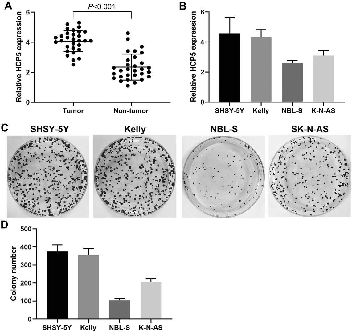 Fig. 1. The expression level of HCP5 in neuroblastoma was up-regulated and related to cell proliferation (Zhu K, Wang L, et al., 2021).
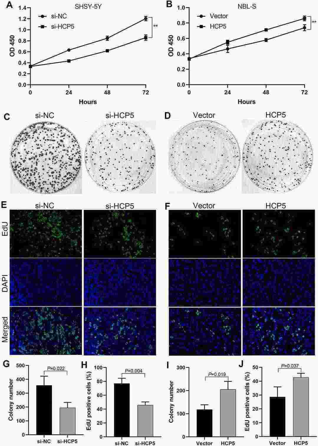 Fig. 2. HCP5 promoted the proliferation of neuroblastoma cells (Zhu K, Wang L, et al., 2021).
PI3K Activation in Neuroblastoma Cells is Predominantly Mediated by p110α but not by p110β or p110δ
Neuroblastoma exhibits high heterogeneity and frequent relapses, challenging conventional treatments. Molecular targeting is seen as a viable alternative. In this study, the role of the PI3K isoform p110α, prevalent in neuroblastoma growth, was studied through in vitro and in vivo experiments.
They utilized the R2: Genomics Analysis and Visualization Platform to examine the expression of class IA PI3K isoforms in neuroblastoma and found that elevated PIK3CA expression correlates with poor prognosis and advanced disease stages. In addition to the differential correlation of class IA PI3K isoforms with neuroblastoma patient outcomes, we tried to determine the isoform dependence of neuroblastoma to PI3Ks. The efficacy of PI3K inhibitors with distinct isoform specificity was compared in neuroblastoma cells (Fig. 3a). The study included cell lines with genetic alterations in MYCN and ALK. Five MYCN-amplified cell lines (LA1-5S, IMR-32, BE(2)C, LAN-5, KELLY) and three non-amplified lines (SH-SY5Y, NBL-S, SK-N-AS) were tested for cytotoxicity by class IA PI3K inhibitors. SH-SY5Y and KELLY have the ALKF1174L mutation, LAN-5 has ALKR1275Q, while LA1-5s lacks ALK expression. We confirmed the expression levels of ALK and MYCN in these lines to understand gene variation. MYCN shows high expression in LAN-5, KELLY, BE(2)C, and IMR-32, and ALK is highly expressed in LAN-5, IMR-32, KELLY, NBL-S, and SH-SY5Y. They found that copanlisib effectively reduced the viability of all tested neuroblastoma cell lines (Fig. 3b). With IC50 values under 1 μM, it was the most effective of the three p110α inhibitors. The pan-PI3K inhibitor pictilisib was also cytotoxic but less effective than copanlisib, hindered by poor solubility in some lines (Fig. 3c). Alpelisib, a p110α-selective inhibitor, reduced cell viability at less than 5 μM (Fig. 3d). Conversely, p110β inhibitor TGX-221 and p110δ inhibitor idelalisib did not affect cell proliferation below 10 μM (Fig. 3e, f), indicating neuroblastoma cells rely on p110α for growth.
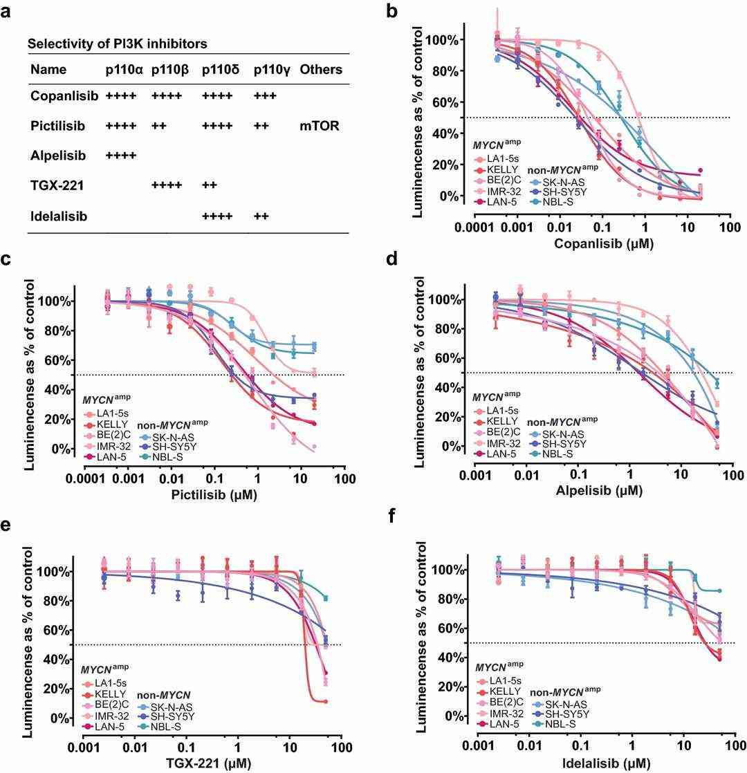 Fig. 3. p110α inhibitors decrease the viability of neuroblastoma cells (Guo Y, Guo D H, et al., 2022).
Ask a Question
Write your own review
- You May Also Need

Description: Subline of GOTO. Protein-free medium adapted. Cell growth is slow.


Description: Established from the adrenal tumor tissue resected after treatment from a 20-month-old boy of European origin with neuroblastoma (stage III) in 1991
Description: established from the bone marrow metastasis of a 1-year-old female patient with adrenal gland neuroblastoma negative

Description: The line is resistant to infection and focus formation by ecotropic MuLV.
Description: Established from a neuroblastoma metastasis at an adrenal site of a 4-year-old white boy in 1986; cells were described to express neuron-specific enolase and synaptophysin (but neither GFAP nor S-100 ...
- Adipose Tissue-Derived Stem Cells
- Human Neurons
- Mouse Probe
- Whole Chromosome Painting Probes
- Hepatic Cells
- Renal Cells
- In Vitro ADME Kits
- Tissue Microarray
- Tissue Blocks
- Tissue Sections
- FFPE Cell Pellet
- Probe
- Centromere Probes
- Telomere Probes
- Satellite Enumeration Probes
- Subtelomere Specific Probes
- Bacterial Probes
- ISH/FISH Probes
- Exosome Isolation Kit
- Human Adult Stem Cells
- Mouse Stem Cells
- iPSCs
- Mouse Embryonic Stem Cells
- iPSC Differentiation Kits
- Mesenchymal Stem Cells
- Immortalized Human Cells
- Immortalized Murine Cells
- Cell Immortalization Kit
- Adipose Cells
- Cardiac Cells
- Dermal Cells
- Epidermal Cells
- Peripheral Blood Mononuclear Cells
- Umbilical Cord Cells
- Monkey Primary Cells
- Mouse Primary Cells
- Breast Tumor Cells
- Colorectal Tumor Cells
- Esophageal Tumor Cells
- Lung Tumor Cells
- Leukemia/Lymphoma/Myeloma Cells
- Ovarian Tumor Cells
- Pancreatic Tumor Cells
- Mouse Tumor Cells