
CAL-85-1
Cat.No.: CSC-C0474
Species: Homo sapiens (Human)
Source: Breast
Morphology: adherent epithelial-like cells growing in monolayers
Culture Properties: monolayer
- Specification
- Background
- Scientific Data
- Q & A
- Customer Review
Immunology: cytokeratin +, cytokeratin-7 +, cytokeratin-8 +, cytokeratin-17 +, cytokeratin-18 +, cytokeratin-19 +, desmin -, endothel -, EpCAM +, GFAP -, neurofilament -,
CAL-85-1 was originally derived in 1990 from a 35-year-old woman who had been diagnosed with relapsing invasive ductal carcinoma of the breast. Cells display an adherent morphology that resembles epithelial-like growth. CAL-85-1 is basal-like and triple-negative (ER/PR-/HER2-) breast adenocarcinoma (TNBC) cell line with TP53 and BRCA2 mutations which are frequently altered in human breast cancer. It has been shown to have an intrinsic multidrug resistance (MDR) phenotype.
CAL-85-1 is frequently utilized in oncology high-throughput screens such as DepMap and CCLE to identify dependencies and druggable targets in TNBC. Additionally, this cell line has been used as a tool to screen novel chemotherapeutics, study DNA damage response in BRCA-deficient cells, and generate more representative 3D culture conditions/or organoids for tumor microenvironment assays.
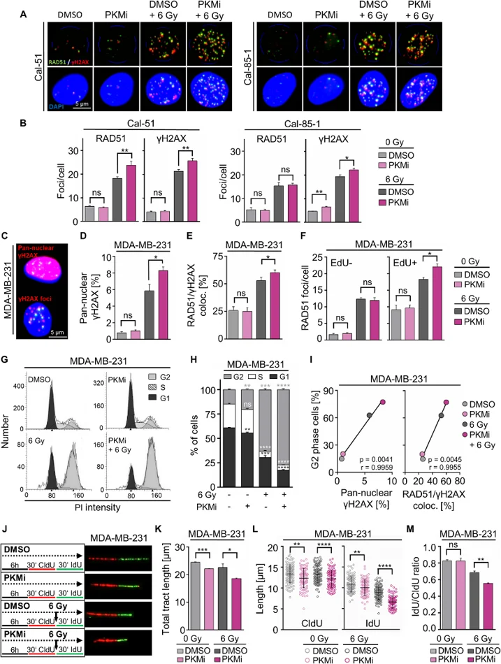
PKM Inhibition Modulates the Radiation Response in TNBC Cells by Regulating DNA Damage Repair, Cell Cycle Progression, and DNA Replication
Metabolic reprogramming contributes to TNBC therapy resistance. Using untargeted metabolomics and a 44-gene interaction network, Matesanz-Sánchez et al. identified radiation-induced metabolic changes and nine radiosensitizing genes by RNAi screening. High PKM expression correlated with poor survival in METABRIC-TNBC; pharmacological PKM inhibition radiosensitized cells through S-phase disruption, reducing DNA synthesis while increasing replication stress and DNA damage at active forks, ultimately prolonging cell cycle arrest.
Mechanistically, combined PKMi/irradiation elevated γH2AX foci in Cal-51 and Cal-85-1 cells (Fig. 1A-B) and increased pan-nuclear γH2AX in MDA-MB-231 and Cal-85-1 cells (Fig. 1C-D), with enhanced RAD51/γH2AX colocalization (Fig. 1E). This damage was replication-dependent, shown by elevated RAD51 foci in EdU-positive MDA-MB-231 cells (Fig. 1F). Significant G2 accumulation occurred in MDA-MB-231 and Cal-85-1 cells (Fig. 1G-H), with G2 arrest correlating with pan-nuclear γH2AX and RAD51/γH2AX colocalization (Fig. 1I).
Ask a Question
Write your own review
- You May Also Need
Description: MCF10DCIS.com is a clonal breast cancer cell line derived from a xenograft originating from premalignant MCF10AT cells that were injected into severe combined immune-deficient mice. MCF10AT is ...

Description: Species: human, Caucasian female 69 years old; Tissue: breast; Tumor: adenocarcinoma; Transfected with ER-pBact, human estrogen receptor cDNA, under control of the human beta actin promotor, ...

Description: Human malignant mesothelioma. CA19-9, CA125, and hyaluronic acid producing. Cell growth is slow.

Description: Histopathology: breast cancerNote: NK (natural killer) cell resistant clone

Description: ZR-75-30 was derived from malignant ascites fluid from a 47-year-old premenopausal Black woman with infiltrating ductal carcinoma.

Description: Species: human - female, 74 years old, CaucasianTumorigenecity: yes, in nude miceIsoenzyme: G6PD, B; PGM3, 1; PGM1, 1; ES-D, 1; Me-2, 0; AK-1, 1; GLO-1, 1Histopathology: carcinoma, ductal
- Adipose Tissue-Derived Stem Cells
- Human Neurons
- Mouse Probe
- Whole Chromosome Painting Probes
- Hepatic Cells
- Renal Cells
- In Vitro ADME Kits
- Tissue Microarray
- Tissue Blocks
- Tissue Sections
- FFPE Cell Pellet
- Probe
- Centromere Probes
- Telomere Probes
- Satellite Enumeration Probes
- Subtelomere Specific Probes
- Bacterial Probes
- ISH/FISH Probes
- Exosome Isolation Kit
- Human Adult Stem Cells
- Mouse Stem Cells
- iPSCs
- Mouse Embryonic Stem Cells
- iPSC Differentiation Kits
- Mesenchymal Stem Cells
- Immortalized Human Cells
- Immortalized Murine Cells
- Cell Immortalization Kit
- Adipose Cells
- Cardiac Cells
- Dermal Cells
- Epidermal Cells
- Peripheral Blood Mononuclear Cells
- Umbilical Cord Cells
- Monkey Primary Cells
- Mouse Primary Cells
- Breast Tumor Cells
- Colorectal Tumor Cells
- Esophageal Tumor Cells
- Lung Tumor Cells
- Leukemia/Lymphoma/Myeloma Cells
- Ovarian Tumor Cells
- Pancreatic Tumor Cells
- Mouse Tumor Cells