
HMMME
Cat.No.: CSC-C6446J
Species: Homo sapiens (Human)
Source: Pleural Effusion
Morphology: epithelial-like
Culture Properties: Adherent cells
- Specification
- Background
- Scientific Data
- Q & A
- Customer Review
Store in liquid nitrogen.
HMMME is a human malignant mesothelioma cell line established from mesothelial tumor tissue. The cells are often used as an in vitro model of malignant mesothelioma, an aggressive cancer that originates from the mesothelial lining of the pleura, peritoneum, or other serosal surfaces. Mesothelioma is a disease primarily associated with asbestos exposure and is marked by rapid progression, local invasiveness, and limited response to treatment.
In culture, HMMME cells typically show adherent growth with epithelial to polygonal morphology, in line with their mesothelial origin. The cells express mesothelioma-associated markers, such as cytokeratins, vimentin, and mesothelial markers like calretinin and mesothelin, consistent with their tumor-derived, epithelial-mesenchymal characteristics. This cell line exhibits strong proliferative capacity and tumor-associated signaling activity, making it a useful tool for mechanistic and translational studies.
HMMME cells are widely used in basic and translational research on mesothelioma pathogenesis, including studies of cell proliferation, apoptosis, invasion, and tumor-microenvironment interactions. The cell line is also utilized for preclinical testing of chemotherapeutic agents, targeted therapies, and other emerging treatment strategies, such as immunomodulatory approaches. As a human cell line with mesothelioma origin and disease relevance, HMMME provides an important experimental platform for advancing the understanding of mesothelioma biology and supporting drug discovery and development efforts.
Risk Prediction for Metastasis of Clear Cell Renal Cell Carcinoma Using Digital Multiplex Ligation-Dependent Probe Amplification
Precise quantification of copy-number alterations (CNAs) in tumor genomes is challenging. Yoshikawa et al. applied digital multiplex ligation-dependent probe amplification (digitalMLPA) for targeted gene copy-number analysis in clear cell renal cell carcinoma (ccRCC) to understand the roles of CNAs in tumor aggressiveness and metastasis.
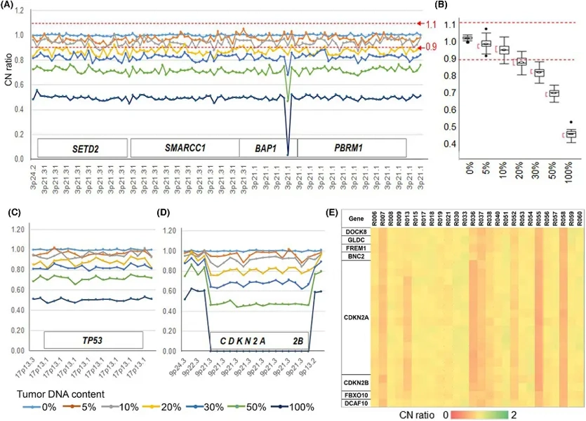
To assess the sensitivity and reproducibility of CNA detection using digitalMLPA, titration DNA experiments were conducted. Tumor cell line DNA from HMMME or H2452, both with losses in 3p21 and 9p21, was mixed with normal cell DNA (HEKn) at proportions ranging from 5% to 100%. Monoallelic losses were detected with high reproducibility at 20% tumor cell line DNA (HMMME) in 80% normal DNA in the 3p21 (Fig. 1A, B) and 17p13 regions (Fig. 1C). Biallelic deletion of the CDKN2A/2B region was detected at 10% tumor cell line DNA (H2452) in 90% normal DNA (Fig. 1D). The precision of the digitalMLPA assay was confirmed by comparing CNA data at BAP1 with data obtained from conventional MLPA (P417-B1 BAP1 probemix) for 34 ccRCCs. The results were essentially identical: 31 of 34 ccRCCs showed 1 allele loss, and the remaining 3 had no CNA.
Ask a Question
Write your own review
- You May Also Need

Description: MCF10DCIS.com is a clonal breast cancer cell line derived from a xenograft originating from premalignant MCF10AT cells that were injected into severe combined immune-deficient mice. MCF10AT is ...

Description: Species: human, Caucasian female 69 years old; Tissue: breast; Tumor: adenocarcinoma; Transfected with ER-pBact, human estrogen receptor cDNA, under control of the human beta actin promotor, ...

Description: Histopathology: breast cancerNote: NK (natural killer) cell resistant clone

Description: ZR-75-30 was derived from malignant ascites fluid from a 47-year-old premenopausal Black woman with infiltrating ductal carcinoma.

Description: Species: human - female, 74 years old, CaucasianTumorigenecity: yes, in nude miceIsoenzyme: G6PD, B; PGM3, 1; PGM1, 1; ES-D, 1; Me-2, 0; AK-1, 1; GLO-1, 1Histopathology: carcinoma, ductal

Description: Species: human, Caucasian female 69 years old; Tissue: breast; Tumor: adenocarcinoma; Transfected with the exon 5 defective variant of human estrogen receptor cDNA, cloned in pZeoSV vector, resistant ...
- Adipose Tissue-Derived Stem Cells
- Human Neurons
- Mouse Probe
- Whole Chromosome Painting Probes
- Hepatic Cells
- Renal Cells
- In Vitro ADME Kits
- Tissue Microarray
- Tissue Blocks
- Tissue Sections
- FFPE Cell Pellet
- Probe
- Centromere Probes
- Telomere Probes
- Satellite Enumeration Probes
- Subtelomere Specific Probes
- Bacterial Probes
- ISH/FISH Probes
- Exosome Isolation Kit
- Human Adult Stem Cells
- Mouse Stem Cells
- iPSCs
- Mouse Embryonic Stem Cells
- iPSC Differentiation Kits
- Mesenchymal Stem Cells
- Immortalized Human Cells
- Immortalized Murine Cells
- Cell Immortalization Kit
- Adipose Cells
- Cardiac Cells
- Dermal Cells
- Epidermal Cells
- Peripheral Blood Mononuclear Cells
- Umbilical Cord Cells
- Monkey Primary Cells
- Mouse Primary Cells
- Breast Tumor Cells
- Colorectal Tumor Cells
- Esophageal Tumor Cells
- Lung Tumor Cells
- Leukemia/Lymphoma/Myeloma Cells
- Ovarian Tumor Cells
- Pancreatic Tumor Cells
- Mouse Tumor Cells