
Sprague-Dawley (SD) Rat Mesenchymal Stem Cells with GFP
- Specification
- Background
- Scientific Data
- Q & A
- Customer Review
Sprague-Dawley (SD) Rat Mesenchymal Stem Cells with GFP are primary bone marrow-derived mesenchymal stem cells (BMSCs) obtained from the femur/tibia of healthy SD rats and stably transduced with a lentiviral vector encoding Green Fluorescent Protein (GFP) for real-time fluorescence tracking. These cells show typical MSC features namely, spindle shaped adherent growth, fibroblast-like form, expression of mesenchymal surface markers (CD29, CD44, and CD90 >70%) and lack of hematopoietic surface markers (CD34, CD45, and CD11b <5%), and the ability to differentiate in vitro into osteoblasts, adipocytes, and chondrocytes in vitro. These are usually cultivated in a basal nutritional medium supplemented with serum and growth factors, under standard conditions (37 °C, 5% CO₂) at a recommended seeding density of 2-4 × 10⁴ cells/cm², and passaged before over-confluence to maintain multipotency (limited to ~5-10 passages).
The integrated GFP reporter enables immediate visualization of cell survival, migration and homing following in vivo transplantation without further labeling. Thus, these GFP-labeled SD rat MSCs are widely used in the preclinical field to study cell tracking, engraftment efficiency, immunomodulatory, and paracrine effects in various rat disease models, including orthotopic bone/cartilage repair, spinal cord or myocardial infarction models, and so on.
RSP Recruits Endogenous Mesenchymal Stem Cells to Rat Ischemic Hindlimbs
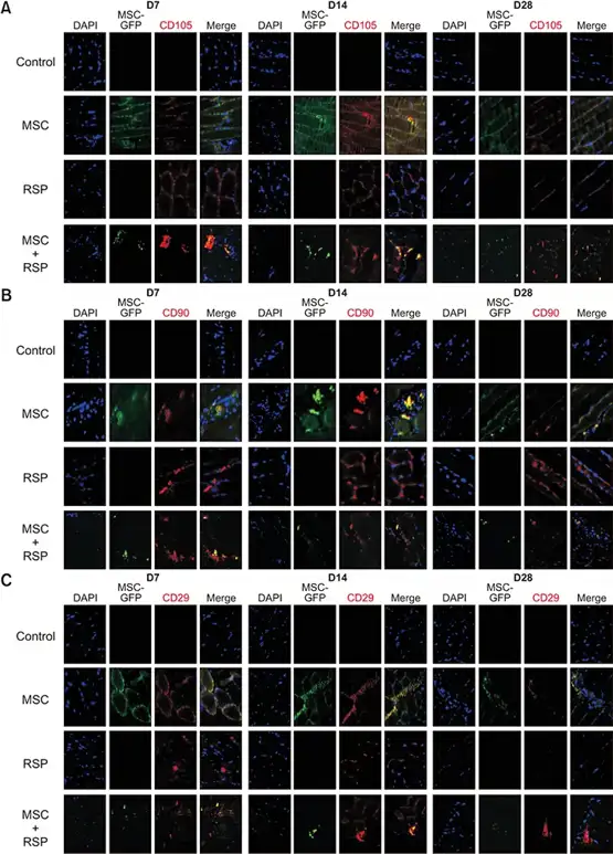
Mesenchymal stem cell (MSC)-based therapies face limitations in critical limb ischemia (CLI) due to poor sustainability. Park et al. evaluated whether a substance P-conjugated scaffold (RSP) could enhance angiogenesis by improving MSC retention and recruitment.
Immunohistochemistry (IHC) tracked MSCs in rat ischemic hindlimbs at 7, 14, and 28 days (Fig. 1). In the MSC-only group, GFP-labeled exogenous cells (green) co-localized with MSC markers (CD105, CD90, CD29; red). The RSP-only group lacked GFP signal but showed persistent endogenous MSC recruitment (red) up to 28 days. In the MSC + RSP group, merged imaging revealed both exogenous MSCs (yellow, co-localization of red and green) and recruited endogenous MSCs (red only). The control group showed no positive staining for any MSC markers. These results demonstrate that RSP sustains exogenous MSC delivery while simultaneously recruiting endogenous MSCs to the ischemic site, independent of ischemia-induced recruitment alone.
Ask a Question
Write your own review
- Adipose Tissue-Derived Stem Cells
- Human Neurons
- Mouse Probe
- Whole Chromosome Painting Probes
- Hepatic Cells
- Renal Cells
- In Vitro ADME Kits
- Tissue Microarray
- Tissue Blocks
- Tissue Sections
- FFPE Cell Pellet
- Probe
- Centromere Probes
- Telomere Probes
- Satellite Enumeration Probes
- Subtelomere Specific Probes
- Bacterial Probes
- ISH/FISH Probes
- Exosome Isolation Kit
- Human Adult Stem Cells
- Mouse Stem Cells
- iPSCs
- Mouse Embryonic Stem Cells
- iPSC Differentiation Kits
- Mesenchymal Stem Cells
- Immortalized Human Cells
- Immortalized Murine Cells
- Cell Immortalization Kit
- Adipose Cells
- Cardiac Cells
- Dermal Cells
- Epidermal Cells
- Peripheral Blood Mononuclear Cells
- Umbilical Cord Cells
- Monkey Primary Cells
- Mouse Primary Cells
- Breast Tumor Cells
- Colorectal Tumor Cells
- Esophageal Tumor Cells
- Lung Tumor Cells
- Leukemia/Lymphoma/Myeloma Cells
- Ovarian Tumor Cells
- Pancreatic Tumor Cells
- Mouse Tumor Cells