
Sprague-Dawley (SD) Rat Mesenchymal Stem Cells
- Specification
- Background
- Scientific Data
- Q & A
- Customer Review
Rat mesenchymal stem cells (rMSCs) are multipotent stromal cells isolated from bone marrow. rMSCs display a characteristic fibroblast-like, spindle-shaped morphology in culture. They form colony-forming unit-fibroblasts (CFU-F) and undergo robust expansion in standard media supplemented with fetal bovine serum, while maintaining stability through multiple passages. Immunophenotypically, rMSCs are positive for CD29, CD44, CD73, CD90, and CD105, and negative for hematopoietic markers CD34, CD45, and CD11b.
A fundamental advantage of rMSCs is their proven trilineage differentiation potential under defined induction conditions. Beyond mesodermal lineages, rMSCs exhibit transdifferentiation toward neural and endothelial phenotypes, underscoring their versatility. The rat model itself confers distinctive translational value: rats are physiologically, metabolically, and anatomically closer to humans than mice, with larger organ dimensions that facilitate surgical manipulation, implantation, and serial blood sampling. rMSCs are therefore extensively employed in preclinical studies modeling myocardial infarction, spinal cord injury, stroke, acute kidney injury, osteoarthritis, and hepatic fibrosis. Their strong immunomodulatory capacity-via paracrine secretion of TGF-β, IL-10, and PGE2-enables allogeneic use without major rejection. Furthermore, rMSCs are readily engineered with lentiviral or CRISPR/Cas9 vectors for gene overexpression or knockdown, and can be labeled with GFP or luciferase for non-invasive tracking. Despite inherent donor heterogeneity, standardized isolation protocols yield reproducible populations, cementing rMSCs as an indispensable ex vivo and in vivo tool for mechanistic interrogation and cell-based therapeutic development.
DMF Preconditioning Enhances Neurotrophic Factor Expression in Rat Bone Marrow Mesenchymal Stem Cells
Stroke is a leading cause of death and disability in adults worldwide. Among various therapeutic strategies, cell-based therapies have gained considerable attention due to their regenerative potential. Enhancing the efficacy of stem cells is crucial for improving therapeutic outcomes. Dimethyl fumarate (DMF) is a drug known to modulate the paracrine effects of stem cells. This study aimed to investigate the effect of different concentrations of DMF on rat bone marrow mesenchymal stem cells (BM-MSCs).
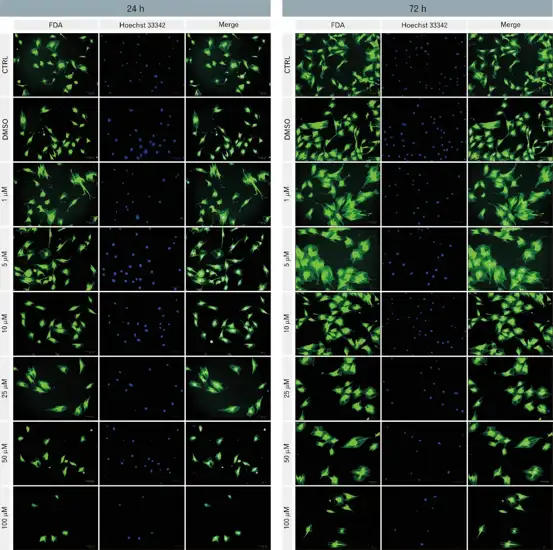
The BM-MSCs viability following treatment with various doses of DMF was assessed using the MTT assay and fluorescein diacetate staining at 24 and 72 hours. After identifying the optimal DMF concentration, BM-MSCs were cultured with selected DMF concentration for 72 hours, and their gene expression profiles of key neurotrophic factors were analyzed via qRT-PCR. The results revealed that 1 µM DMF was the optimal concentration for enhancing BM-MSC viability. Treatment with this dose significantly upregulated the expression of brain-derived neurotrophic factor (BDNF), nerve growth factor (NGF), and neurotrophin-3 (NT-3), highlighting their potential in promoting neuronal support and regeneration. In contrast, the transcript level of glial-derived neurotrophic factor (GDNF) was significantly reduced, suggesting a selective regulatory effect of DMF on neurotrophic pathways. These findings shed light on the therapeutic promise of DMF in modulating neurotrophic factor expression in BM-MSCs, offering novel insights into its application in regenerative medicine for neurodegenerative conditions.

Ask a Question
Write your own review
- Adipose Tissue-Derived Stem Cells
- Human Neurons
- Mouse Probe
- Whole Chromosome Painting Probes
- Hepatic Cells
- Renal Cells
- In Vitro ADME Kits
- Tissue Microarray
- Tissue Blocks
- Tissue Sections
- FFPE Cell Pellet
- Probe
- Centromere Probes
- Telomere Probes
- Satellite Enumeration Probes
- Subtelomere Specific Probes
- Bacterial Probes
- ISH/FISH Probes
- Exosome Isolation Kit
- Human Adult Stem Cells
- Mouse Stem Cells
- iPSCs
- Mouse Embryonic Stem Cells
- iPSC Differentiation Kits
- Mesenchymal Stem Cells
- Immortalized Human Cells
- Immortalized Murine Cells
- Cell Immortalization Kit
- Adipose Cells
- Cardiac Cells
- Dermal Cells
- Epidermal Cells
- Peripheral Blood Mononuclear Cells
- Umbilical Cord Cells
- Monkey Primary Cells
- Mouse Primary Cells
- Breast Tumor Cells
- Colorectal Tumor Cells
- Esophageal Tumor Cells
- Lung Tumor Cells
- Leukemia/Lymphoma/Myeloma Cells
- Ovarian Tumor Cells
- Pancreatic Tumor Cells
- Mouse Tumor Cells