
Human iPSC-derived renal proximal tubular cells
- Specification
- Background
- Scientific Data
- Q & A
- Customer Review
hiPSC-derived renal proximal tubular cells (hiPSC-derived PTCs/PTLs) are functional epithelial-like cells that are created by guided differentiation of human induced pluripotent stem cells (hiPSCs) into the renal proximal tubule lineage. The cells show significant morphological and physiological properties of natural proximal tubular epithelial cells (PTECs) and serve as a viable in vitro model for kidney research and translational toxicology.
hiPSC-derived proximal tubular cells have conventional epithelial shape and express typical proximal tubule markers, including megalin (LRP2), cubilin, AQP1 and brush border-type transport proteins. Functionally, they exhibit albumin endocytosis, the activity of transporters (OATs, OCTs) and polarized epithelial characteristics which are required to imitate renal reabsorption and xenobiotic processing in vivo.
More recently, these cells have been shown to respond in a physiologically realistic manner to known nephrotoxins such as cisplatin, cyclosporine A and aminoglycosides, supporting their application in nephrotoxicity screening and medication safety assessment. Furthermore, proximal tubular models created from hiPSCs are being included into kidney organoid systems and microfluidic organ-on-chip platforms, allowing for increased maturation, enhanced transporter expression and more faithful modelling of renal physiology.
In summary, human iPSC-derived renal proximal tubular cells provide a robust and scalable human-relevant model to study renal development, drug-induced kidney injury, transporter biology and disease mechanisms, bridging the gap between the conventional cell lines and primary human renal epithelial cells.
Characterization of hiPSC Differentiation into Proximal Tubule Epithelial Cells
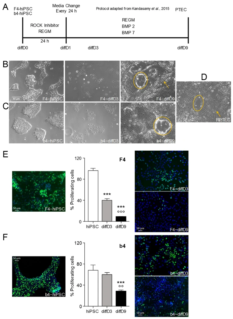
Mboni-Johnston et al. investigated the sensitivity of human induced pluripotent stem cells (hiPSC) and their differentiated progeny to nephrotoxins. Dermal fibroblast-derived hiPSC lines (F4 and b4) were differentiated into proximal tubular epithelial-like cells (PTELC) following the protocol outlined in Figure 1A.
During differentiation, both cell lines transitioned morphologically from a cobblestone to a spindle-like shape by day 9, forming domes and tubule-like structures (Fig. 1B-D). Proliferation rates, assessed by EdU incorporation, dropped sharply in F4 cells from nearly 100% to approximately 10% by day 9 (Fig. 1E), whereas b4 cells exhibited a slower decline, retaining about 30% proliferation at day 9 (Fig. 1F).
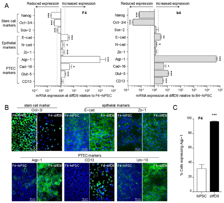
Gene expression analysis revealed significant downregulation of stem cell markers (NANOG, Oct-3/4) in both lines after nine days, with Sox-2significantly reduced only in F4 (Fig. 2A). Conversely, epithelial markers like E-cadherin increased. Among four proximal tubule markers, all were significantly elevated in differentiated b4 cells, while only three (Aqp-1, Cad-16, Glut-5) increased significantly in F4 cells, with CD13mRNA unchanged. Immunocytochemistry confirmed these changes at the protein level (Fig. 2B), revealing discrepancies between mRNA and protein expression for CD13 and Zo-1. Flow cytometry demonstrated that nearly all differentiated F4 cells expressed the PTEC marker Aquaporin-1 by day 9, a threefold increase over undifferentiated controls (Fig. 2C).
Ask a Question
Write your own review
- Adipose Tissue-Derived Stem Cells
- Human Neurons
- Mouse Probe
- Whole Chromosome Painting Probes
- Hepatic Cells
- Renal Cells
- In Vitro ADME Kits
- Tissue Microarray
- Tissue Blocks
- Tissue Sections
- FFPE Cell Pellet
- Probe
- Centromere Probes
- Telomere Probes
- Satellite Enumeration Probes
- Subtelomere Specific Probes
- Bacterial Probes
- ISH/FISH Probes
- Exosome Isolation Kit
- Human Adult Stem Cells
- Mouse Stem Cells
- iPSCs
- Mouse Embryonic Stem Cells
- iPSC Differentiation Kits
- Mesenchymal Stem Cells
- Immortalized Human Cells
- Immortalized Murine Cells
- Cell Immortalization Kit
- Adipose Cells
- Cardiac Cells
- Dermal Cells
- Epidermal Cells
- Peripheral Blood Mononuclear Cells
- Umbilical Cord Cells
- Monkey Primary Cells
- Mouse Primary Cells
- Breast Tumor Cells
- Colorectal Tumor Cells
- Esophageal Tumor Cells
- Lung Tumor Cells
- Leukemia/Lymphoma/Myeloma Cells
- Ovarian Tumor Cells
- Pancreatic Tumor Cells
- Mouse Tumor Cells