Canine Bone Marrow Mesenchymal Stem Cells
- Specification
- Background
- Scientific Data
- Q & A
- Customer Review
Never can cryopreserved cells be kept at -20 °C.
Canine bone marrow mesenchymal stem cells (cBM-MSCs) are multipotent adult stem cells residing in the bone marrow stroma. They are defined by their plastic adherence, fibroblast-like morphology, expression of characteristic surface markers (CD44+, CD90+, CD105+, and negativity for hematopoietic markers such as CD34- and CD45-), and trilineage differentiation capacity into osteocytes, chondrocytes, and adipocytes under defined inductive conditions.
cBM-MSCs have gained significant attention in both veterinary medicine and translational research. Unlike human MSCs, canine models offer the advantage of studying spontaneous, naturally occurring diseases - including osteoarthritis, intervertebral disc degeneration, and immune-mediated disorders - that closely mimic human pathologies. Key advantages of cBM-MSCs include: (1) robust expansion ex vivo - they can be isolated from small aspirates and expanded to clinically relevant numbers while retaining multipotency; (2) immunomodulatory properties - they suppress T-cell proliferation and secrete anti-inflammatory cytokines (IL-10, TGF-β), supporting their use in allogeneic transplantation; (3) functional cryopreservation - they maintain viability and potency after freeze-thaw cycles; (4) osteochondral competence - they reliably undergo chondrogenesis and osteogenesis, making them ideal for cartilage and bone repair studies; (5) species-relevant preclinical model - canine studies provide translationally valuable data for human regenerative medicine, particularly for large animal models and clinical trials.
Collectively, cBM-MSCs represent a reproducible, versatile, and clinically relevant cell source for investigating musculoskeletal regeneration, inflammatory diseases, and cell-based therapies, bridging the gap between rodent models and human applications.
Unraveling the Osteogenic Potentials of PCL/HA Scaffolds in Canine Mesenchymal Stem Cells
Currently, the development of composite scaffolds has emerged as an attractive approach to meet the criteria of ideal scaffolds in bone tissue engineering (BTE). Recently, the incorporation of polycaprolactone (PCL) with hydroxyapatite (HA) has been developed as one of the suitable substitutes for BTE applications owing to their promising osteogenic properties.
In this study, a three-dimensional (3D) scaffold composed of PCL integrated with HA (PCL/HA) was prepared and assessed for its ability to support osteogenesis in vitro. Furthermore, this scaffold was evaluated explicitly for its efficacy in promoting the proliferation and osteogenic differentiation of canine bone marrow-derived mesenchymal stem cells (cBM-MSCs) to fill the knowledge gap regarding the use of composite scaffolds for BTE in the veterinary orthopedics field.
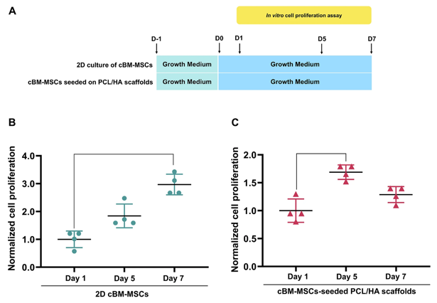
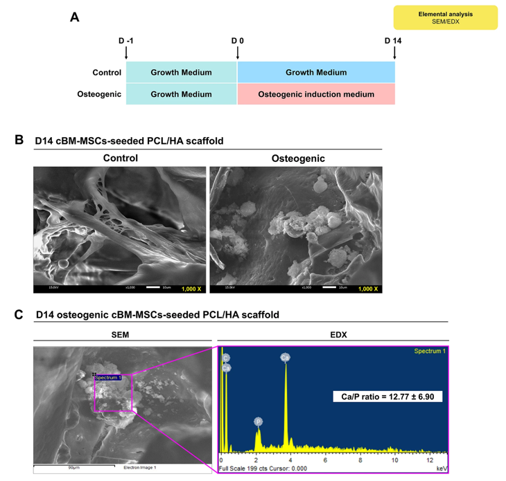
The findings indicate that the PCL/HA scaffolds substantially supported the proliferation of cBM-MSCs. Notably, the group subjected to osteogenic induction exhibited a markedly upregulated expression of the osteogenic gene osterix (OSX) compared to the control group. Additionally, the construction of 3D scaffold constructs with differentiated cells and an extracellular matrix (ECM) was successfully imaged using scanning electron microscopy. Elemental analysis using a scanning electron microscope coupled with energy-dispersive X-ray spectroscopy confirmed that these constructs possessed the mineral content of bone-like compositions, particularly the presence of calcium and phosphorus.
Ask a Question
Write your own review
- You May Also Need
- Adipose Tissue-Derived Stem Cells
- Human Neurons
- Mouse Probe
- Whole Chromosome Painting Probes
- Hepatic Cells
- Renal Cells
- In Vitro ADME Kits
- Tissue Microarray
- Tissue Blocks
- Tissue Sections
- FFPE Cell Pellet
- Probe
- Centromere Probes
- Telomere Probes
- Satellite Enumeration Probes
- Subtelomere Specific Probes
- Bacterial Probes
- ISH/FISH Probes
- Exosome Isolation Kit
- Human Adult Stem Cells
- Mouse Stem Cells
- iPSCs
- Mouse Embryonic Stem Cells
- iPSC Differentiation Kits
- Mesenchymal Stem Cells
- Immortalized Human Cells
- Immortalized Murine Cells
- Cell Immortalization Kit
- Adipose Cells
- Cardiac Cells
- Dermal Cells
- Epidermal Cells
- Peripheral Blood Mononuclear Cells
- Umbilical Cord Cells
- Monkey Primary Cells
- Mouse Primary Cells
- Breast Tumor Cells
- Colorectal Tumor Cells
- Esophageal Tumor Cells
- Lung Tumor Cells
- Leukemia/Lymphoma/Myeloma Cells
- Ovarian Tumor Cells
- Pancreatic Tumor Cells
- Mouse Tumor Cells