Animal Primary Cells
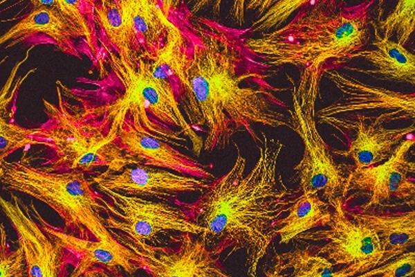
The animal cell model is an alternative for in vivo studying the biological functions of cells. As models for human systems, researchers can use animal cells to examine a wide range of disease mechanisms and evaluate novel treatments before applying the results of these investigations to humans. Animal cell culture has found use in diverse areas, from basic to advanced research.
- Studying basic cell biology, cell cycle mechanisms, specialized cellular functions, cell-cell and cell-matrix interactions
- Toxicity testing to study the effects of new drugs
- Gene therapy for replacing non-functional genes with functional gene-carrying cells
- Production of vaccines, monoclonal antibodies, and pharmaceutical drugs
- Production of viruses for use in vaccine production
Creative Bioarray's animal primary cells are isolated from normal or diseased animal tissues. They are rigorously quality tested to meet our high standard and specifications.
- Over 500 different lots available
- Isolated from both healthy and diseased animals
- Cryopreserved immediately after isolation
- Stored in frozen vials
Search our available specified cell types to find primary cell solutions for your research.
Filters Clear all filters
Species
- Bovine (22)
- Cat (45)
- Chicken (11)
- Chinchilla (1)
- Dog (118)
- Fish (1)
- Fruitfly (1)
- Goat (46)
- Guinea Pig (8)
- Hamster (94)
- Horse (1)
- Human (3)
- Minipig (2)
- Monkey (129)
- Mouse (872)
- Pig (110)
- Rabbit (249)
- Rat (323)
- Sheep (2)
- Squirrel (1)
- Turkey (1)
Source
- Adipose (23)
- Adrenal Gland (7)
- Airway (1)
- Anus (3)
- Aorta (70)
- Artery (133)
- Bile Duct (8)
- Bladder (41)
- Blood (36)
- Bone (10)
- Bone Marrow (76)
- Brain (127)
- Breast (54)
- Bronchus (23)
- Cartilage (23)
- Cervix (5)
- Chorion (3)
- Choroid (6)
- Colon (53)
- Conjunctiva (7)
- Cornea (25)
- Dermis (68)
- Diaphragm (3)
- Ear (12)
- Embryo (22)
- Endometrium (9)
- Epidermis (7)
- Epididymis (3)
- Esophagus (31)
- Eye (75)
- Gallbladder (3)
- Gingiva (18)
- Hair Follicle (10)
- Heart (58)
- Intestine (132)
- Kidney (120)
- Lens (3)
- Liver (83)
- Lung (137)
- Lymph Node (25)
- Mesentery (15)
- Nose (4)
- Olfactory Bulb (1)
- Oral Cavity (9)
- Ovary (62)
- Oviduct (7)
- Pancreas (60)
- Pancreatic Duct (3)
- Pancreatic Islet (10)
- Parathyroid Gland (4)
- Penis (7)
- Periodontal Ligament (3)
- Periodontium (21)
- Peripheral Blood (23)
- Peritoneal Cavity (12)
- Placenta (18)
- Prostate (54)
- Pudenda (2)
- Rectum (2)
- Retina (29)
- Salivary Gland (3)
- Sclera (3)
- Skeletal Muscle (27)
- Skin (77)
- Small Intestine (51)
- Spinal Cord (9)
- Spleen (70)
- Stomach (34)
- Synovium (5)
- Tendon (7)
- Testis (12)
- Thymus (48)
- Thyroid (30)
- Tongue (5)
- Trabecular Meshwork (2)
- Trachea (40)
- Umbilical Cord (6)
- Ureter (10)
- Urethra (3)
- Uterus (54)
- Vein (74)
Cell Type
- Adipocyte (4)
- Astrocyte (25)
- B Cell (8)
- Basal Cell (3)
- Beta Cell (3)
- Cardiomyocyte (16)
- CD34+ Cell (5)
- Cholangiocyte (8)
- Chondrocyte (15)
- Dendritic Cell (11)
- Endothelial Cell (555)
- Endothelial Progenitor Cell (7)
- Epithelial Cell (432)
- Fibroblast (334)
- Glial Cell (44)
- Goblet Cell (1)
- Granule Cell (1)
- Granulocyte (9)
- Granulosa Cell (1)
- Hepatic Stellate Cell (6)
- Hepatocyte (14)
- Interstitial Cell (9)
- Keratinocyte (10)
- Keratocyte (2)
- Kupffer Cell (7)
- Leydig Cell (3)
- Lymphocyte (29)
- Macrophage (27)
- Mast Cell (3)
- Melanocyte (1)
- Meningeal Cell (4)
- Mesangial Cell (8)
- Mesothelial Cell (3)
- Microglia (6)
- Microvascular Cell (247)
- Monocyte (9)
- Mononuclear Cell (12)
- Myofibroblast (3)
- Myosatellite Cell (1)
- Neuron (43)
- Neutrophil (8)
- NK Cell (6)
- Oligodendrocyte (3)
- Oligodendrocyte Progenitor Cell (1)
- Osteoblast (5)
- Osteoclast (2)
- Osteocyte (3)
- Pancreatic Stellate Cell (3)
- Pericyte (10)
- Podocyte (4)
- Preadipocyte (11)
- Progenitor Cell (11)
- Red Blood Cell (11)
- Retinal Ganglion Cell (3)
- Satellite Cell (1)
- Schwann Cell (3)
- Sertoli Cell (4)
- Skeletal Muscle Cell (9)
- Smooth Muscle Cell (193)
- Spermatogonium (3)
- Stromal Cell (25)
- Synoviocyte (5)
- T Cell (11)
- Tenocyte (7)
- Trabecular Meshwork Cell (2)
- Trophoblast (3)
Disease
- Breast Cancer (5)
- Cancer (23)
- Cervical Cancer (2)
- Colon Cancer (5)
- Diabetes (75)
- Lung Cancer (6)
- Normal (1942)
- Pancreatic Cancer (2)
- Prostate Cancer (3)

Description: The gingiva is the pink-colored keratinized mucosa that surrounds the teeth. Gingival Fibroblasts ...

Description: The umbilical vein is a blood vessel present during fetal development that carries oxygenated blood ...

Description: The chorion is one of the embryonic membranous structures that encloses the fetus and the amnion. ...

Description: The renal arteries are large blood vessels that carry blood from your heart to kidneys. The kidney ...

Description: The esophagus is the muscular tube that carries food and liquids from mouth to the stomach. ...

Description: The gastric wall has four layers: mucosa, submucosa, muscularis and serosa. Fibroblasts are the ...

Description: The small intestine is located in the abdomen, the upper end is connected to the stomach through ...

Description: The small intestine is located in the abdomen, the upper end is connected to the stomach through ...

Description: The small intestine is located in the abdomen, the upper end is connected to the stomach through ...

Description: The colon wall has four layers: mucosa (epithelium, lamina propria, muscularis mucosa), submucosa ...
Description: Intrahepatic bile duct epithelial cells account for about 5% of the total number of hepatocytes, ...

Description: The extrahepatic bile ducts are part of a network of ducts that carry bile from the liver and ...

Description: The liver is an essential organ that has many functions in the body, including making proteins and ...

Description: Kupffer cells are a critical component of the mononuclear phagocytic system and are central to both ...

Description: The aortic valve is a valve in the heart of humans and most other animals, located between the left ...

Description: The uterus is the organ that produces menstruation and gestation of the fetus, which is located in ...

Description: The femoral head is the most proximal portion of the femur and is supported by the femoral neck. ...

Description: The thoracic aorta is located in the chest cavity and gives off arteries that branch to the ...