
Human Esophageal Smooth Muscle Cells (HESMC)
Cat.No.: CSC-7781W
Species: Human
Source: Esophagus
Cell Type: Smooth Muscle Cell
- Specification
- Background
- Scientific Data
- Q & A
- Customer Review
Human esophageal smooth muscle cells (HESMC) are primary smooth muscle cells isolated from the human esophageal muscularis propria, typically from the lower esophagus. They retain a contractile, donor-specific phenotype and express canonical smooth muscle markers, including α-smooth muscle actin, SM22α, calponin, desmin, h-caldesmon, and smooth muscle myosin heavy chain. Functionally, HESMC exhibit agonist-induced calcium signaling and contraction, proliferation, migration, and extracellular matrix synthesis. They respond to acetylcholine, KCl, endothelin-1, angiotensin II, serotonin, histamine, and inflammatory cytokines, reflecting native esophageal smooth muscle physiology.
Compared with immortalized smooth muscle lines and animal-derived cells, HESMC better preserve native receptor expression, contractile apparatus, and donor genetic background, improving translational relevance. Because they are human and site-specific, they reduce xenogeneic variability and support personalized studies of esophageal motility disorders and fibrotic remodeling. They are widely used to study esophageal motility, achalasia, gastroesophageal reflux disease, eosinophilic esophagitis, esophageal fibrosis, and tissue-engineered esophageal constructs. They also support drug screening for prokinetic, antispasmodic, anti-inflammatory, and anti-fibrotic agents.
Chronic Hypoxia-Induced Reprogramming of Smooth Muscle Cells
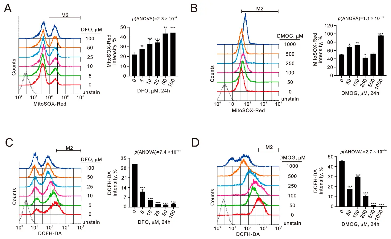
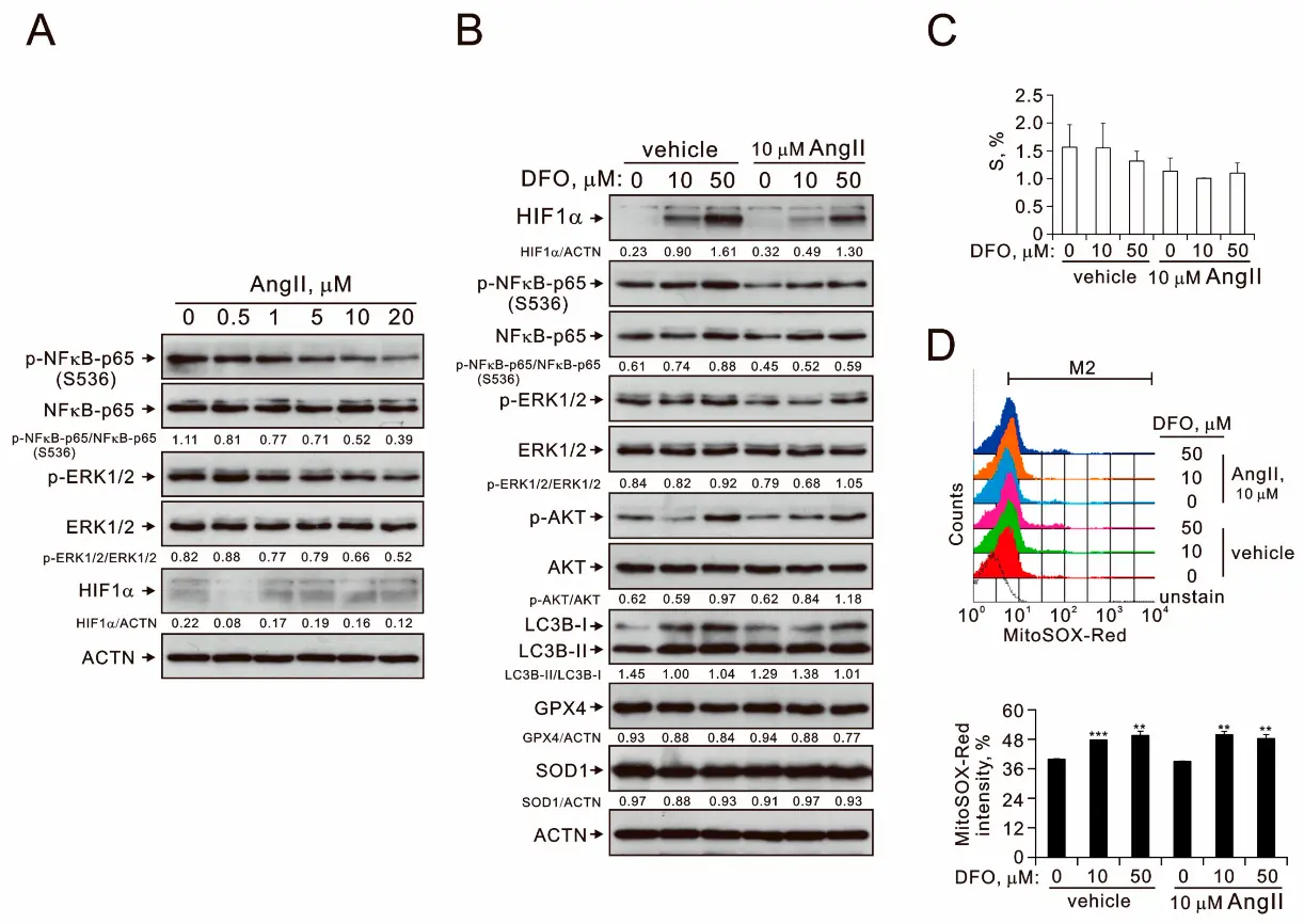
Chronic hypoxia promotes a shift in smooth muscle cells from a quiescent, contractile state to a highly proliferative phenotype, contributing to persistent vascular remodeling. This study examines primary human esophageal smooth muscle cells (HEsSMCs) exposed to hypoxia mimics, desferoxamine (DFO) and dimethyloxallyl glycine (DMOG), and their interaction with angiotensin II (AngII).
The findings indicate that both DFO and DMOG stabilize hypoxia-inducible factor-1 alpha proteins at different time intervals, revealing distinct cell cycle profiles and varied protein responses. DFO was shown to increase mitochondrial reactive oxygen species (ROS) while simultaneously suppressing cytosolic ROS, suggesting the involvement of multiple pathways beyond those activated by hypoxic stress. Flow cytometry analysis revealed that DFO triggers late-stage apoptosis, along with increases in overall hypoxia, ROS, autophagy, lipid peroxidation, and mitochondrial membrane depolarization in HEsSMCs. Furthermore, AngII was observed to inhibit the activation of nuclear factor-κB induced by DFO.
Ask a Question
Write your own review
Description: HEMEC from Creative Bioarray are isolated from human esophageal tissue. HEMEC are cryopreserved at passage one and delivered frozen. Each vial contains >5 x 10^5 cells in 1 ml volume. HEMEC are ...
Description: The human esophagus is lined by a non-keratinizing, moist stratified squamous epithelium whose apical cell membranes and intercellular junctional complexes combine to produce an effective ...
Description: The HEF cells from Creative Bioarray are isolated from human esophageal tissue. HEF are cryopreserved at passage one and delivered frozen. Each vial contains >5 x 10^5 cells in 1 ml volume. HEF are ...