
Human Esophageal Fibroblasts (HEF)
Cat.No.: CSC-7782W
Species: Human
Source: Esophagus
Cell Type: Fibroblast
- Specification
- Background
- Scientific Data
- Q & A
- Customer Review
The esophagus is the digestive tract located between the pharynx and the stomach. The structure of the mammalian esophagus is divided into four layers from the inside to the outside: the mucosa, submucosa, muscular layer, and the outer membrane. Esophageal fibroblasts, which line the lamina propria of the upper and lower esophagus, are mesenchymal cells derived from the embryonic mesoderm. Fibroblasts secrete a non-rigid extracellular matrix that is both rich in type I and/or type III collagen. They are responsible for the synthesis of extracellular matrix in connective tissues and play a central role in wound healing. They have been extensively used in various cellular and molecular studies, as they are one of the cell types that are easy to grow in culture. In addition, their durability makes them suitable for a variety of manipulations, from gene transfection to microinjection.
Human Esophageal Fibroblasts (HEF) from Creative Bioarray are isolated from human esophageal tissue. HEF are cryopreserved at passage one and delivered frozen. Each vial contains more than 5 x 10⁵ cells. HEF are characterized by immunofluorescence with antibody specific to fibronectin. HEF are negative for HIV-1, HBV, HCV, mycoplasma, bacteria, yeast and fungi. Creative Bioarray also provides optimized media and reagents for the growth of human esophageal fibroblasts, which are quality tested together and guaranteed to give maximum performance as a global solution for in vitro HEF culture.
Anti-Fibrogenic Effect of Umbilical Cord–Derived Mesenchymal Stem Cell–Conditioned Media in Human Esophageal Fibroblasts
Umbilical cord-derived mesenchymal stem cells (UC-MSCs) are known to mitigate fibrosis in various organs. However, the potential effects of UC-MSCs on human esophageal fibrosis remain underexplored. This study investigated the anti-fibrogenic properties and mechanisms of UC-MSC-derived conditioned media (UC-MSC-CM) on human esophageal fibroblasts (HEFs). HEFs were treated with TGF-β1 and then cultured with UC-MSC-CM, and the expression levels of extracellular matrix (ECM) components, RhoA, myocardin related transcription factor A (MRTF-A), serum response factor (SRF), Yes-associated protein (YAP), and transcriptional coactivator with PDZ-binding motif (TAZ) were measured.
UC-MSC-CM suppressed TGF-β1-induced fibrogenic activation in HEFs, as evidenced by the downregulation of ECM. UC-MSC-CM diminished the expression of RhoA, MRTF-A, and SRF triggered by TGF-β1. In TGF-β1-stimulated HEFs, UC-MSC-CM decreased the nuclear localization of MRTF-A and YAP. Additionally, UC-MSC-CM diminished the TGF-β1-induced nuclear expressions of YAP and TAZ, while concurrently enhancing the cytoplasmic presence of phosphorylated YAP. Furthermore, UC-MSC-CM reduced TGF-β1-induced phosphorylation of Smad2.
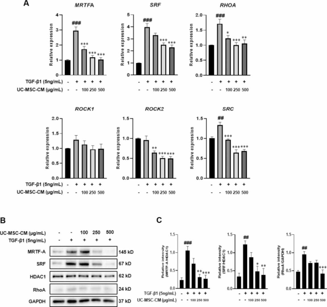 Fig. 1. UC-MSC-CM inhibits Rho/MRTF/SRF signaling in HEFs (Choi, Yoon Jeong, et al. 2024).
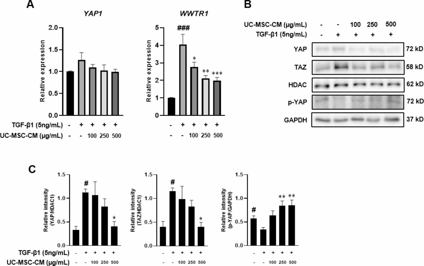 Fig. 2. UC-MSC-CM inhibits YAP/TAZ signaling in HEFs (Choi, Yoon Jeong, et al. 2024).
Ask a Question
Write your own review

Description: HEMEC from Creative Bioarray are isolated from human esophageal tissue. HEMEC are cryopreserved at passage one and delivered frozen. Each vial contains >5 x 10^5 cells in 1 ml volume. HEMEC are ...
Description: The human esophagus is lined by a non-keratinizing, moist stratified squamous epithelium whose apical cell membranes and intercellular junctional complexes combine to produce an effective ...
Description: Smooth muscle is responsible for the contractility of hollow organs, such as blood vessels, the gastrointestinal tract, the bladder, and the uterus. Its structure differs greatly from that of ...