IHC Protocols Using Anti-GFP Antibodies
GUIDELINE
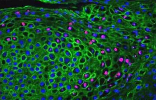
The green fluorescent protein (GFP) from the jellyfish Aequorea victoria is a versatile marker for monitoring physiological processes, visualizing protein localization, and detecting transgenic expression. We offer the anti-GFP antibody as a rabbit polyclonal, monoclonal, or IgG fraction, two mouse monoclonal antibodies, and a chicken IgY fraction. All six anti-GFP antibodies are suited for the detection of native GFP, GFP variants, and most GFP fusion proteins by western blot analysis while the rabbit and mouse antibodies are also useful for immunoprecipitation.
METHODS
- Remove the media from the cells grown on coverslips. Rinse the cells twice for 1 minute each in D-PBS.
- Fix the cells in fixative solution (4% formaldehyde in PBS) for 30 minutes at room temperature with gentle agitation in the dark. Remove the solution.
- Wash the cells twice in PBS for 1 minute each with gentle agitation. Remove the PBS.
- Permeabilize the specimen with permeabilization solution for 5 minutes at room temperature with gentle agitation in the dark. Remove the solution.
- Wash the cells twice in PBS for 1 minute each with gentle agitation. Remove the PBS.
- Add blocking solution (5% normal goat serum in PBS, pH 7.4) to the cells. Incubate for 1 hour at room temperature with gentle agitation. Remove the solution.
- Wash the cells twice in PBS for 1 minute each with gentle agitation.
- Prepare a 1:400 dilution of anti-GFP chicken antibody in PBS to obtain a final antibody concentration of 5.0 µg/mL.
- Remove the PBS and add the diluted primary antibody solution to the cells. Incubate for 1 hour at room temperature with gentle agitation. Remove the solution.
Wash the cells twice in PBS for 1 minute each with gentle agitation. - Prepare the appropriate conjugated secondary antibody in PBS.
- Remove the PBS and add the diluted secondary antibody solution to the cells. Incubate for 1 hour at room temperature with gentle agitation. Remove the solution.
- Wash the cells twice in PBS for 2 minutes each with gentle agitation. After the final wash, add PBS to the sample. The sample is now ready for imaging and detection using an appropriate method of choice.
Creative Bioarray Relevant Recommendations
- Creative Bioarray offers a comprehensive IHC service from project design, and marker selection to image completion and data analysis. We are dedicated to satisfying every customer and assisting them to achieve their specific research goals.
- We provide a large number of fluorescent products, which are the most trusted products available. Our team is always committed to providing customers with high-quality products and services.
| Cat. No. | Product Name |
| FDIR-D0031 | GFP-LC3 fluorescent lentivirus |
| FDIR-D0032 | GFP-p62 fluorescent lentivirus |
| FDIR-D0043 | GFP-Vimentin fluorescent lentivirus |
| FDIR-D0049 | GFP-Rad51 fluorescent lentivirus |
| FDIR-D0051 | GFP-Tubulin fluorescent lentivirus |
| FDIR-D0053 | GFP-HMGB1 fluorescent lentivirus |
| FDIR-D0035 | Paxillin-GFP fluorescent lentivirus |
| FDIR-D0037 | α-actin-GFP fluorescent lentivirus |
| FDIR-D0039 | β-actin-GFP fluorescent lentivirus |
| FDIR-D0041 | EB3-GFP fluorescent lentivirus |
| FDIR-D0045 | PSD95-GFP fluorescent lentivirus |
| FDIR-D0047 | Histone H2B-GFP fluorescent lentivirus |
NOTES
- Ensure proper permeabilization of the tissue if intracellular GFP expression needs to be detected. This can be achieved through the use of detergents such as Triton X-100 or saponin.
- Consider counterstaining the tissue with nuclear dyes or other cellular markers to provide context to the GFP expression within the tissue.
- Include appropriate positive and negative controls in the IHC experiment to validate the specificity of the anti-GFP antibody and the overall staining procedure.