
Why Your Cells Won't Detach? And How to Fix It?
Mastering cell detachment is equal parts science and art. Whether you are struggling with stubborn cells that refuse to let go of the plate, or watching cell viability plummet after a harsh harvest, detachment issues can quickly derail weeks of careful culture work. Achieving high viability and consistent single-cell suspensions requires looking beyond standard protocol timers to understand what is happening at the cell surface.
Quick Diagnostic Flowchart
When a harvest goes off-script, identifying the root cause quickly saves your culture. Start by matching your culture’s behavior to one of three common scenarios:
Pathway A: Stubborn Attachment or Aggregation
- What you see: Cells remain firmly anchored after incubation, or blow off in large, sticky sheets that will not disperse.
- Likely culprits: Dense extracellular matrix (ECM) accumulation, residual divalent cations, or degraded enzyme activity.
Pathway B: Low Viability and High Debris
- What you see: Rapid detachment followed by low post-passage survival, visible cellular fragments, or poor re-attachment.
- Likely culprits: Over-incubation, excessive enzyme concentration, delayed neutralization, or rough pipetting.
Pathway C: Unintended Spontaneous Detachment
- What you see: Cells lift or float on their own during normal incubation without any dissociation reagents.
- Likely culprits: Overconfluency, hidden mycoplasma or bacterial contamination, media pH swings, or stress-induced apoptosis.
The Biology Behind the Bond
To fix detachment problems, it helps to understand how cells cling to their environment—and why breaking those bonds can sometimes go wrong.
Integrins and Extracellular Matrix (ECM) Dynamics
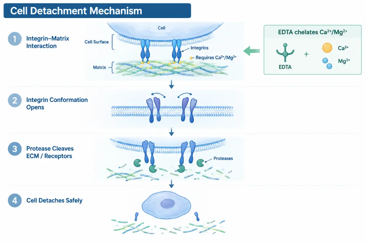
Adherent cells anchor themselves to plastic surfaces using integrin receptors that grip matrix proteins like fibronectin, collagen, and laminin. These integrin bridges rely heavily on divalent cations (Ca2+ and Mg2+) to maintain their structural grip. If calcium or magnesium remains in your wash buffer, these anchors stay rigid, leaving digestive enzymes working against an uphill battle.
Trypsin Kinetics and the Danger of DNA Nets
Trypsin works by cutting peptide bonds at lysine and arginine residues. While effective, it is a blunt instrument. Prolonged exposure strips crucial membrane proteins and damages delicate cell membranes. When cells lyse, they release strands of sticky genomic DNA. This free DNA acts like a net, trapping healthy single cells into tight, irreversible clumps that pipetting will only make worse.
Overconfluency and Tight Junctions
When cultures grow beyond 80–90% confluency, cells begin linking tightly to each other through cadherins and desmosomes while laying down a thick ECM layer. This dense physical network blocks dissociation reagents from penetrating down to the cell-substrate interface.
Practical Troubleshooting Guide
Issue 1: Cells Refuse to Come Off
Serum Traces Neutralizing the Enzyme: Fetal bovine serum (FBS) contains strong protease inhibitors like α1-antitrypsin that shut trypsin down instantly.
The Fix: Always wash the cell sheet twice with Ca2+/Mg2+-free phosphate-buffered saline (DPBS−/−) before adding your dissociation agent.
High Matrix Producers: Primary fibroblasts and smooth muscle cells form exceptionally tough ECM networks.
The Fix: Use a formulation paired with EDTA (e.g., 0.25% Trypsin / 1 mM EDTA). EDTA strips the stabilizing cations, exposing the protein backbone to enzymatic cleavage. For heavy matrices, consider adding collagenase or dispase.
Cold Reagents: Trypsin activity drops off significantly below 37°C.
The Fix: Pre-warm your reagents and return the vessel to the 37°C incubator during digestion rather than leaving it on the benchtop.
Issue 2: Poor Viability and Clumping Post-Harvest
Harsh Handling: Excessive, rapid pipetting to break up non-dissociated cell sheets creates shear stress that shreds cell membranes.
The Fix: Ditch violent pipetting. Gently tap the side of the flask instead, and use wide-orifice tips to protect delicate membranes.
Extracellular DNA Accumulation: Over-digestion releases DNA that binds living cells together.
The Fix: Add DNase I (20–50 μg/mL) to your cell suspension to dissolve the DNA web without harming live cells. Always neutralize trypsin immediately with serum-rich media or a dedicated inhibitor at a 1:1 ratio.
Issue 3: Loss of Downstream Surface Markers
Proteolytic Cleavage: Standard trypsinization destroys surface antigens like CD markers needed for flow cytometry or single-cell RNA sequencing.
The Fix: Switch to gentle, non-enzymatic dissociation buffers or recombinant enzymes designed to preserve membrane integrity.
Tailored Detachment Protocols
Different cell types require a tailored approach. Here is a quick reference guide for common cell models:
| Cell Category | Examples | Recommended Agent | Optimal Conditions | Pro-Tip |
|---|---|---|---|---|
| Standard Adherent | HeLa, HEK293, NIH3T3 | 0.25% Trypsin-EDTA | 37°C, 1–3 min | Check under the scope; stop as soon as 80% of cells round up. |
| High ECM / Strong Adhesion | Primary Fibroblasts, Smooth Muscle Cells | 0.25% Trypsin-EDTA + Collagenase | 37°C, 5–10 min | Double wash with DPBS-/-; pre-warm all solutions to 37°C. |
| Sensitive & Stem Cells | hPSCs, iPSCs, Neural Progenitors | Recombinant Protease / Accutase | 37°C, 3–5 min | Supplement with ROCK inhibitor post-harvest to prevent apoptosis. |
| Marker-Sensitive Lines | Immune Subsets, Flow Samples | Non-Enzymatic Chelating Buffer | 37°C or RT, 5–15 min | Ideal for preserving surface receptors and antigens. |
| Semi-Adherent / Macrophages | RAW 264.7, Microglia | Cold PBS + Gentle Cell Scraper | Ice-cold, 5–10 min | Avoid standard enzymes; scrape gently at a 45° angle. |
Golden Rules for Everyday Culture Success
- Never Skip Divalent-Free Washes: A thorough DPBS−/− rinse removes serum inhibitors and loosens cation-dependent anchors.
- Trust Your Eyes, Not Just the Clock: Always monitor detachment under an inverted microscope. Stop digestion the moment cells round up and start lifting.
- Keep Incubation Short: Minimize enzyme exposure to the bare minimum needed for detachment.
- Manage Passage Density: Subculture before reaching 80% confluency to keep cell-matrix networks manageable.
- Screen for Mycoplasma: Unexplained cell detachment or poor adhesion is often an early warning sign of silent mycoplasma contamination.
Frequently Asked Questions
Q1: Why are my cells still attached after 10 minutes in trypsin?
In most cases, residual serum was left behind to neutralize the trypsin, or the enzyme degraded from too many freeze-thaw cycles. Wash thoroughly with DPBS−/−, use fresh trypsin-EDTA, and consider adding collagenase if working with high-matrix cells.
Q2: How exactly does EDTA help with cell detachment?
EDTA chelates Ca2+ and Mg2+ ions. Because integrins and cadherins require these cations to hold their structure and cling to the plastic, removing them opens up the protein conformation so enzymes can do their job faster.
Q3: How do I get rid of persistent cell clumps after centrifugation?
Clumping usually stems from sticky DNA released by dead cells. Shorten your incubation times, handle cells gently, and add DNase I (20–50 μg/mL) to your resuspension buffer to clear the DNA nets cleanly.