
Scaling Organoid Screening: A Hands-On Guide for 3D Drug Discovery
Introduction: Moving Beyond the "Proof-of-Concept" Trap
If you have ever worked with patient-derived organoids (PDOs), you probably know that magical feeling when you look under the microscope and see a gorgeous, textbook-perfect 3D mini-organ thriving in a dome of Matrigel. In an academic or exploratory setting, that is a massive win.
But if you are sitting on a preclinical drug discovery or screening team, you quickly run into a humbling reality: growing a pretty organoid is a world away from screening 10,000 compounds against it.
Traditional 2D cell cultures are fast, cheap, and play nicely with high-throughput screening (HTS) robots—but they are basically biologically blind. Animal models, on the other hand, give you the whole biological picture, but their metabolism and genes often don't match human responses, which is why so many promising animal-tested drugs crash and burn in human clinical trials.
Organoids are the ultimate bridge. They mimic real human tissues and disease microenvironments like nothing else. But the bottleneck has officially moved. The question is no longer "Can we grow them?" It is "How on earth do we optimize, standardize, and scale them so they don't break our screening pipeline?"
Diagnosing the Breakdown: Why Traditional HTS Breaks Down in 3D
Before we talk about fixing things, let's look at why standard HTS protocols throw a temper tantrum the moment you try to use 3D models.
The Matrigel Mess
Most labs start out using Matrigel (basement membrane extract harvested from mouse tumors). Sure, cells love it, but for industrial screening, it is a nightmare:
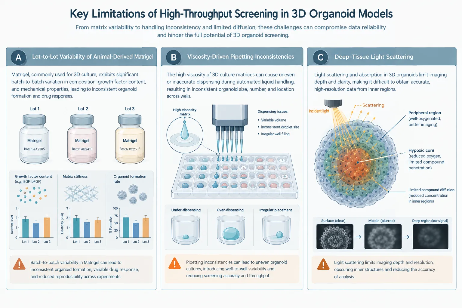
- Lot-to-lot chaos: Because it comes from animals, protein concentrations and mechanical stiffness change from batch to batch. That hidden background noise will ruin your IC50 curves every single time.
- Temperature tantrums: Matrigel polymerizes if it even thinks about warming up. Pipetting it manually leads to clogged tips, uneven droplets, and messy multi-well plates.
The Viscosity Barrier
In a 2D screen, your liquid handler handles cell media like water. Try pumping a viscous 3D hydrogel through a standard robotic nozzle, and you will get wildly uneven cell seeding densities and massive well-to-well variations that ruin your statistical window.
The Diffusion Brick Wall
In living human tissues, blood vessels take care of oxygen and nutrients. In a static 3D organoid that grows larger than 200 micrometers, passive diffusion has to do all the heavy lifting. The outer cells hog everything, the core goes necrotic, and test drugs struggle to penetrate deep inside. If a drug "fails" to work, is it because the cells are resistant, or because the drug couldn't swim through the matrix? Often, it's the latter.
Core Strategy I: Matrix Engineering and Defined Microenvironments
If you want industrial-grade reproducibility, you have to break up with animal-derived extracts and move toward smart, engineered materials.
Switching to Synthetic Hydrogels
Modern screening teams are shifting toward poly(ethylene glycol) (PEG) hydrogels and peptide-functionalized matrices.
- Tune the stiffness: Different tissues like different environments. If you are screening for a liver fibrosis drug, you need a stiff matrix. If you are looking at brain tumor organoids, you need something super soft and compliant. Synthetic hydrogels let you dial in the exact stiffness you want.
- Add the right hooks: By adding specific adhesion peptides (like RGD motifs) into the synthetic backbone, you give cells the exact chemical signals they need to survive and polarize without needing mystery animal proteins.
Strict Pre-Screening QC (Garbage In, Garbage Out)
You cannot just dump a random slurry of different-sized organoids into a 384-well plate and expect clean data.
- Size control: Use automated strainers, gentle enzymatic steps, or microfluidic droplet gadgets to keep your organoids strictly uniform—ideally right between 100 and 150 micrometers in diameter.
- Viability checkpoints: Run an automated image-based viability check. If a batch is sitting below 90% live cells, don't waste your compounds on it.
Core Strategy II: Automation and 384-Well Plate Integration
Scaling up from a manual 96-well plate to a high-throughput 384-well or 1536-well format means your team will burn out fast unless you let the robots take the wheel—carefully.
Acoustic Non-Contact Dispensing (AND)
Delicate 3D organoids and hydrogels hate being jabbed by metal pins or squeezed through standard pipette tips. That is why top-tier screening facilities use acoustic non-contact dispensers. They use focused sound waves to blast tiny, nanoliter-sized droplets of your test compounds straight into the assay wells. No touching, no mechanical stress, zero cross-contamination.
Redefining Your Dosing Clock
Small molecules and antibodies take their sweet time diffusing through a 3D extracellular matrix compared to a flat monolayer.
- Give it time: If your 2D assay calls for a 48-hour drug exposure window, be prepared to extend that to 72, 96, or even 144 hours for 3D organoids. Just make sure your automated workflow includes scheduled media and compound refreshes to keep things fresh.
Core Strategy III: Advanced Readouts and High-Content Imaging
If you rely on old-school bulk assays like CellTiter-Glo 3D to give you a single luminescence number at the end of the run, you are flying blind. You won't know if a drop in signal is because the drug killed the cells, stopped them from growing, or if the core just suffocated.
Optical Clearing for Deep-Tissue Imaging
Big organoids scatter light like crazy, turning confocal microscopes into expensive flashlights.
- Clear the view: Use specialized optical clearing reagents that match the refractive index of your cells to the surrounding media. Suddenly, light passes straight through, letting you take gorgeous z-stack images through the entire thickness of a 200+ micrometer organoid.
AI-Driven Phenotypic Profiling
High-content imaging generates an absolute mountain of multidimensional image data. Don't try to look at it by hand.
- Let ai do the heavy lifting: Modern pipelines use machine learning to automatically find organoids in wells, track whether they are shrinking or swelling, measure nuclear changes, and spot dying cells in real time. Combine this with single-cell RNA sequencing on selected wells, and you get a complete picture of how a drug is working.
Validation Framework and Quality Metrics
Before you pitch your shiny new organoid screen to the project steering committee, you have to prove it's statistically bulletproof.
Chasing a Great Z-Factor
In HTS, the Z' factor is your report card. It measures how cleanly your positive and negative controls are separated.
- The golden rule: Your 3D multi-well assay needs to consistently hit a Z' factor greater than 0.5. If your biological noise is washing out your signal, you need to tighten up your plate-to-plate consistency before running real compounds.
Bench-to-Bedside Validation
The ultimate test of credibility is retrospective validation. Take a panel of drugs with known clinical trial outcomes, run them through your organoid assay against matching patient tumor models, and see if the results line up. If your organoid shrinkage and viability data accurately mirror real clinical responses, you've got a winner.
Conclusion
Optimizing an organoid drug screening workflow isn't about throwing money at the flashiest lab automation on the market. It comes down to a practical checklist:
- Lock down your materials: Ditch unpredictable animal extracts for tunable synthetic matrices and strict size control.
- Automate smartly: Use acoustic dispensing to protect your delicate 3D structures.
- Upgrade your eyes: Use optical clearing and AI imaging so you actually see what's happening inside the organoid.
- Demand statistical rigor: Guard that Z-factor like your job depends on it.
Get those pieces right, and you'll successfully turn organoids from a finicky academic science project into an industrial-grade engine that actually predicts what works in humans.
Creative Bioarray Relevant Recommendations
| Products & Services | Description |
|---|---|
| 3D Spheroid Platform for Drug Development | Creative Bioarray provides advanced 3D spheroid models designed for researchers aiming to explore cell behavior in a more realistic environment. |
| Organoid Platform for Drug Development | Creative Bioarray presents a state-of-the-art Organoid Platform designed to transform drug development and biomedical research. By leveraging advanced organoid and organoid-immune co-culture models, this platform provides highly accurate and predictive solutions that closely replicate human biology. |
| Organ-on-a-Chip Platform for Drug Development | Creative Bioarray focuses on developing specific human disease models and leveraging our organ-on-a-chip platform to identify new therapeutics and clinical biomarkers, promote drug development, and create novel drug delivery systems. |