
How to Choose the Right Positive and Negative Controls for IHC
Immunohistochemistry (IHC) can produce beautiful images, but a visually convincing stain is not automatically a reliable result. A brown or red signal may reflect true target expression, but it can also result from poor antibody specificity, inadequate blocking, tissue-related artifacts, or an issue in the detection system.
That is why controls are not optional extras in IHC. They are the evidence that tells you whether your staining run worked—and whether your experimental samples can be interpreted with confidence.
This guide explains how to choose and use positive controls, negative controls, internal controls, and no-primary controls in a practical IHC workflow.
Why IHC Controls Matter
An IHC result should answer two questions:
Did the staining method work?
Is the observed signal specific to the target?
Controls help answer both questions, but no single control can answer everything.
For example, a strong positive control confirms that the antibody and detection chemistry can generate a signal under your protocol. However, it does not prove that every brown area in your experimental tissue is specific. Likewise, a clean no-primary control suggests that the secondary detection system is not producing obvious background, but it does not prove that the primary antibody binds only to the intended antigen.
A robust IHC assay therefore uses a combination of controls. At minimum, most routine assays should include:
- An external positive control;
- An internal positive control when available;
- A negative tissue control;
- A no-primary antibody control when troubleshooting or validating a new protocol.
The exact mix depends on the assay purpose, tissue type, target abundance, antibody validation status, and whether the staining is being used for research, biomarker development, or diagnostic decision-making.
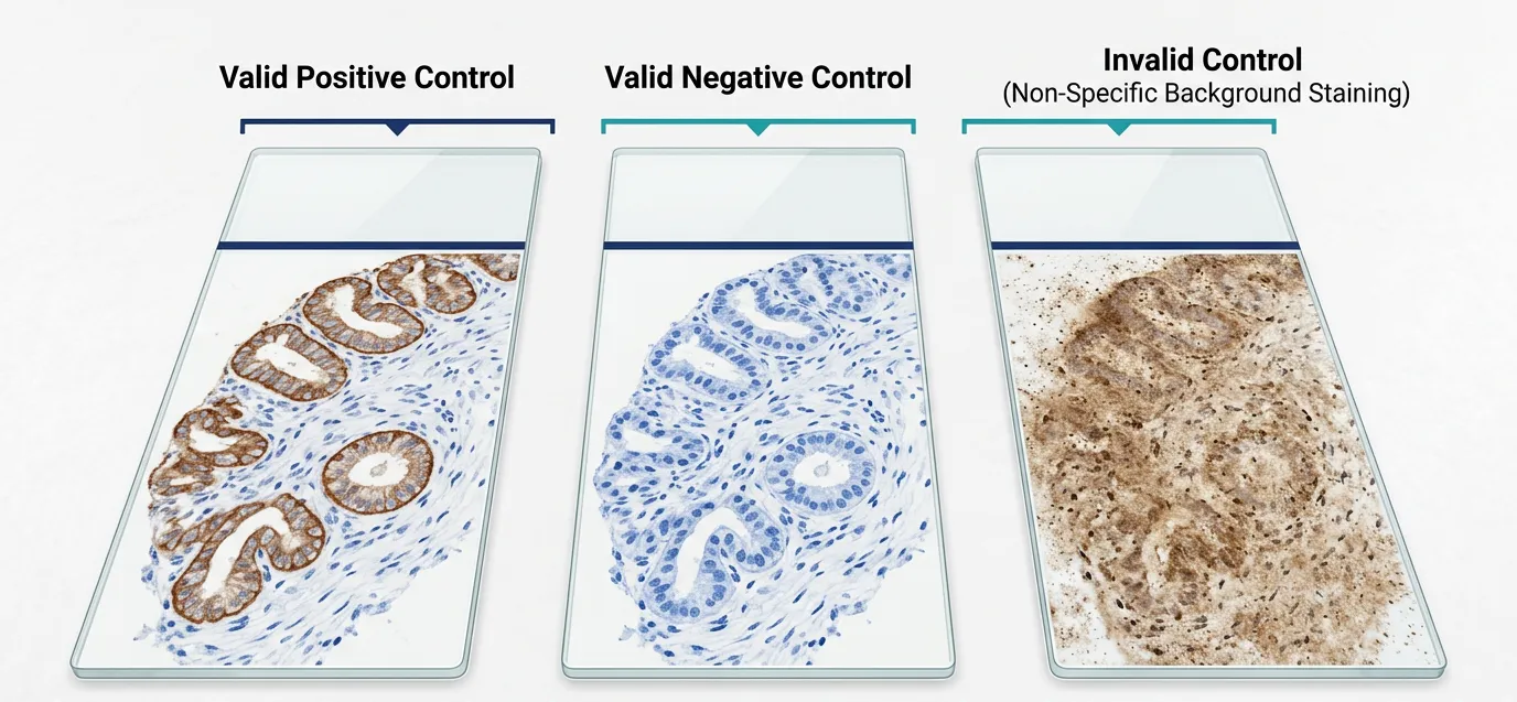 Fig. 1. Valid and invalid IHC control patterns.Positive controls should show expected target
staining, negative controls should remain clean, and diffuse staining may indicate an invalid run.
The Four Main Types of IHC Controls
| Control type | Main purpose | What it can tell you | Main limitation |
|---|---|---|---|
| External positive control | Confirms the assay can detect the target | Antibody, retrieval, detection, and chromogen can produce expected staining | Does not reflect fixation quality of every test sample |
| Internal positive control | Confirms staining performance within the same section | The tissue section retained detectable antigen and the run worked locally | Not available for every target or sample |
| Negative tissue control | Evaluates whether staining appears in tissue expected to lack the target | Helps identify inappropriate tissue distribution or cross-reactivity | "Negative" tissue may still express low levels of the protein |
| No-primary antibody control | Detects background from the secondary system, endogenous enzymes, biotin, or chromogen | Helps identify non-primary-antibody background | Does not establish primary antibody specificity |
Understanding these differences is important. One of the most common IHC mistakes is treating a single control as proof that the whole assay is valid.
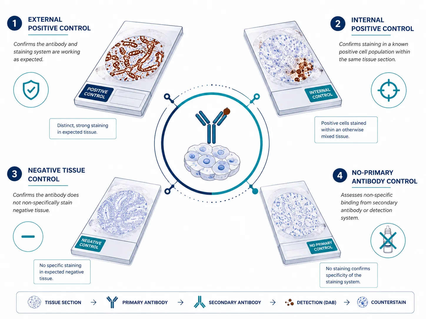 Fig. 2. Four essential control types in IHC. External positive, internal positive, negative
tissue, and no-primary controls address different aspects of assay performance and specificity.
Choosing an External Positive Control
An external positive control is a separate tissue section known to express the target protein. It should be stained alongside your experimental samples using the same protocol.
What makes a good positive control tissue?
A useful positive control should have:
- Well-documented expression of the target;
- Consistent and preferably moderate-to-strong expression;
- Clear expected cellular localization;
- Reliable tissue morphology;
- Similar fixation, processing, and embedding conditions to test samples.
Expression databases, published literature, antibody datasheets, pathology atlases, and prior internal validation data can help identify candidate control tissues.
Avoid relying only on an extremely strong positive control
A tissue with very high target expression is useful for confirming that an antibody can work. However, it may not be ideal for optimizing sensitivity or assessing subtle background.
Very strong expression can hide technical weaknesses. An antibody concentration that looks acceptable in a highly positive tissue may generate excessive background in a low-expression research sample.
Whenever possible, include a control with low-to-moderate expression during assay development. This makes it easier to optimize the signal-to-background ratio and determine whether your method can distinguish true weak positivity from noise.
Match processing conditions whenever possible
The best external control is processed in a way that resembles the experimental specimens.
For FFPE IHC, try to match:
- Fixative type;
- Fixation duration;
- Tissue processing schedule;
- Paraffin embedding conditions;
- Section thickness;
- Antigen retrieval method.
A control tissue fixed for 12 hours may stain well even if your experimental tissue was over-fixed for several days. In that situation, the positive control confirms that the staining reagents are active, but it does not prove that your test tissue retained the antigen.
Internal Positive Controls: The Most Informative Control When Available
An internal positive control is a cell population or tissue structure within the same experimental section that is expected to express the target.
Examples may include:
- Endothelial cells;
- Lymphocytes;
- Smooth muscle cells;
- Normal epithelial cells adjacent to a tumor;
- Stromal cells;
- Resident immune cells.
Internal controls are valuable because they experience the same fixation, sectioning, antigen retrieval, antibody incubation, detection chemistry, and chromogen development as the tissue you are evaluating.
If an internal positive control stains as expected while the target population is negative, that negative result becomes more credible. In contrast, if the internal control fails to stain, it may indicate compromised antigen preservation, unsuitable retrieval conditions, or a staining run failure.
Important limitation
Do not assume a cell type is an internal positive control without evidence. Target expression can vary by tissue site, disease state, species, treatment status, and fixation quality. Confirm the expected expression pattern before using it as a reference.
Choosing a Negative Tissue Control
A negative tissue control is a tissue known—or strongly expected—to lack the target antigen under the conditions being studied.
How to select a negative tissue
Look for tissue with:
- Little or no reported expression of the target;
- Similar tissue composition to the test tissue when possible;
- Similar fixation and processing conditions;
- Minimal natural pigment or endogenous enzyme activity that could confuse interpretation.
A practical caution: negative does not always mean zero expression
Many proteins are expressed at low levels across multiple tissues. A "negative" tissue may show weak physiological staining, especially with sensitive polymer detection systems.
Therefore, interpret negative tissue controls in context. The question is not always "Is there absolutely no color?" but rather:
Is the staining pattern biologically plausible, localized appropriately, and clearly different from the expected positive pattern?
The No-Primary Antibody Control
In a no-primary antibody control, the primary antibody is omitted while all other steps remain unchanged. his control is especially useful during assay development, when optimizing a new antibody, or when unexpected background appears.
- A positive no-primary control may indicate:
- Endogenous peroxidase activity in HRP-DAB IHC;
- Endogenous biotin in avidin-biotin systems;
- Non-specific secondary antibody or polymer binding;
- Excessive chromogen development;
- Inadequate washing;
- Tissue pigment or precipitated reagent.
A clean no-primary control is reassuring, but it does not prove primary antibody specificity. It only suggests that major non-primary sources of background are under control.
For a detailed troubleshooting workflow, link this section to your article on Why Is My IHC Background Too High?
What About Isotype Controls?
Isotype controls use an antibody of the same species and immunoglobulin class as the primary antibody, but without specificity for the target antigen.
They can sometimes help assess non-specific binding associated with antibody isotype or Fc interactions. However, they are often overused or misinterpreted.
An isotype control does not reliably prove that your primary antibody is specific. It may have different affinity, formulation, glycosylation, concentration, and tissue behavior from the target-specific antibody.
Use isotype controls selectively, particularly when Fc receptor-mediated binding is a concern. For most IHC assays, a combination of appropriate positive tissue, negative tissue, no-primary controls, and independent antibody validation is more informative.
Interpreting Control Results
Before interpreting your experimental samples, assess the controls first.
| Control outcome | Likely interpretation | Can test samples be interpreted? |
|---|---|---|
| Positive control positive; negative controls clean | Assay is performing as expected | Yes, if staining pattern is biologically plausible |
| Positive control negative | Antibody, retrieval, detection, or tissue processing may have failed | No; repeat or troubleshoot |
| No-primary control positive | Detection-related background or endogenous tissue activity is present | Not until the cause is resolved |
| Negative tissue shows unexpected staining | Possible cross-reactivity, low-level expression, or non-specific binding | Interpret cautiously and validate further |
| Internal control positive; test cells negative | Negative result may be credible | Usually yes, if biology supports it |
| Internal control negative; external control positive | Test sample may have poor antigen preservation or inadequate retrieval | Usually no; assess sample quality |
A failed control does not necessarily mean every sample result is wrong, but it means the run lacks the evidence needed for confident interpretation.
Controls for FFPE, Frozen Tissue, and Multiplex IHC
FFPE tissue
For FFPE IHC, controls should ideally share similar fixation and processing conditions with the test samples. Antigen retrieval is especially important, since fixation-related epitope masking can vary between blocks.
Include controls that help distinguish true antigen loss from staining failure. Internal controls are particularly valuable in old archival blocks or variably fixed specimens.
Frozen tissue
Frozen sections may preserve some antigens better than FFPE tissue, but they can retain higher endogenous enzyme activity and may be more vulnerable to tissue damage. Include controls processed with the same fixation and permeabilization conditions.
Multiplex IHC
Multiplex IHC requires additional control layers:
- Single-plex staining for each marker;
- Single-color controls for each chromogen or fluorophore;
- Omission controls for individual antibodies;
- Sequential staining controls to test cross-reactivity;
- Spectral or channel controls in fluorescence workflows.
Never assume that markers validated individually will behave identically when combined in a multiplex panel.
Common IHC Control Mistakes
Avoid these frequent problems:
1. Using only a positive control.
A positive slide shows that the assay can generate signal, not that every signal is specific.
2. Choosing an overly strong positive tissue.
Strong controls can conceal poor sensitivity or excessive background.
3. Using controls processed differently from test samples.
This limits their ability to reveal fixation- or processing-related failure.
4. Treating a no-primary control as a specificity control.
It evaluates detection-related background, not the specificity of the primary antibody.
5. Ignoring internal controls.
When available, they provide direct evidence from the same section.
6. Using an assumed negative tissue without verifying expression data.
Low-level physiological expression may be mistaken for non-specific staining.
7. Continuing interpretation after a control failure.
If key controls fail, repeat or troubleshoot before reporting conclusions.
Creative Bioarray Relevant Recommendations
| Products & Services | Description |
|---|---|
| Immunohistochemistry (IHC), Immunofluorescence (IF) Service | Creative Bioarray offers a comprehensive IHC service from project design, marker selection to image completion and data analysis. |
| Histology Services | Creative Bioarray offers tissue processing, embedding, sectioning, and staining, along with set of histological examination services. |