
How to Identify and Troubleshoot Cell Morphology Changes
Cell morphology can reveal changes in cellular behavior, but a change in shape does not always indicate a problem. Learn what drives common morphological changes, how to interpret them, and when additional testing may be needed.

What Drives Changes in Cell Morphology?
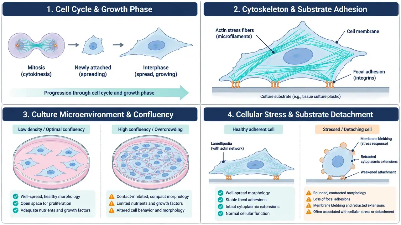
Cell shape is influenced by the cell cycle, cytoskeletal organization, adhesion, and the surrounding culture environment. The same visible change can arise from different biological processes.
- Cell Cycle and Biological State
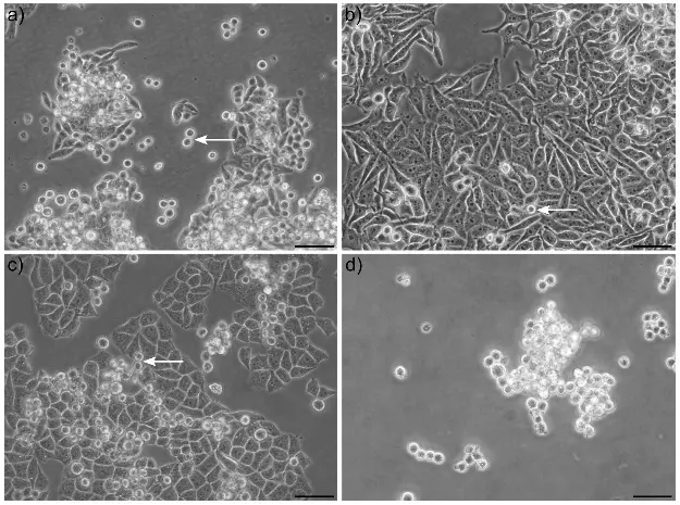
Cells may round up during mitosis and spread again after division. Differentiation can produce cell type-specific shapes, while senescent cells may become enlarged and flattened.
What to check
Compare the observed shape with the expected behavior of the cell type, growth pattern, and relevant phenotype markers.
- Cytoskeleton and Cell Adhesion
The actin cytoskeleton and adhesion structures help cells maintain their shape and attach to the culture surface. Changes in these systems can alter spreading, elongation, contractility, and attachment.
What to check
Review surface coatings, handling procedures, and treatment history. Use cytoskeletal or adhesion assays when the mechanism is relevant.
- Culture Conditions and Microenvironment
Seeding density, confluence, medium composition, serum, substrate properties, and matrix stiffness can influence cell spreading and interactions. Morphological differences may therefore reflect environmental responses rather than intrinsic cell damage.
What to check
Compare culture density, medium and reagent lots, culture vessels, and recent changes in handling or incubation conditions.
- Cellular Stress and Damage
Adverse culture conditions, compound exposure, and contamination may cause rounding, shrinkage, irregular shapes, detachment, or increased debris. These features overlap with normal cellular processes and cannot identify a specific mechanism on their own.
What to check
Assess viability, proliferation, and contamination risk according to the suspected cause and experimental context.
Normal Variation or a Warning Sign?
A single morphological feature rarely provides a definitive answer. Timing, distribution, persistence, and accompanying changes can help narrow down the possibilities.
Observation: Cells become rounded
Possible normal variation
Cells may round up during mitosis and subsequently reattach or spread. Rounding may also result from reduced adhesion or cellular injury.
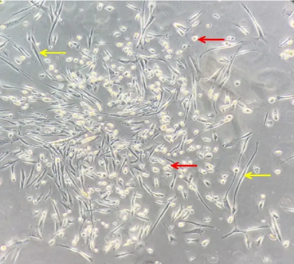
Look for: timing, attachment, progression of the change, and viability.
Observation: Cells become more elongated
Context-dependent
Elongation may reflect cell type-specific morphology, cytoskeletal remodeling, or a change in phenotype.
Look for: historical images, culture density, and phenotype-specific markers if needed.
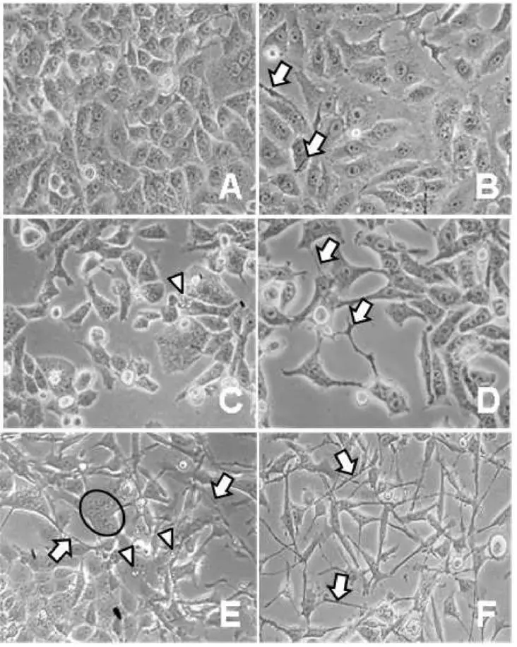
Observation: Cells become larger and flatter
Context-dependent
This may accompany cellular growth or senescence-associated changes, but other processes can produce similar appearances.
Look for: proliferation trends and appropriate markers or functional measurements.
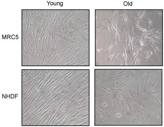
Observation: Many cells detach
Investigate promptly
Potential causes include altered adhesion, handling-related damage, unsuitable culture conditions, cytotoxicity, or contamination.
Look for: recent culture events, viability, medium conditions, and contamination risk.

Quick Reference: What Should You Check Next?
| Observed change | Initial checks | Possible follow-up |
|---|---|---|
| Reduced cell spreading | Confluence, surface, medium, handling | Adhesion or cytoskeletal assessment |
| Slower growth with normal morphology | Cell counts, passage history, culture records | Proliferation and contamination testing |
| Increased cell detachment | Recent handling, medium, treatment | Viability and contamination tests |
| Suspected differentiation | Expected morphology and culture conditions | Phenotype-specific markers and functional assays |
| Persistent batch-to-batch differences | Cell source, passage number, culture procedures | Cell identity and quality-control testing |
These are investigation pathways, not diagnostic rules. Select follow-up tests according to the cell model and the question being investigated.
How to Investigate Unexpected Changes
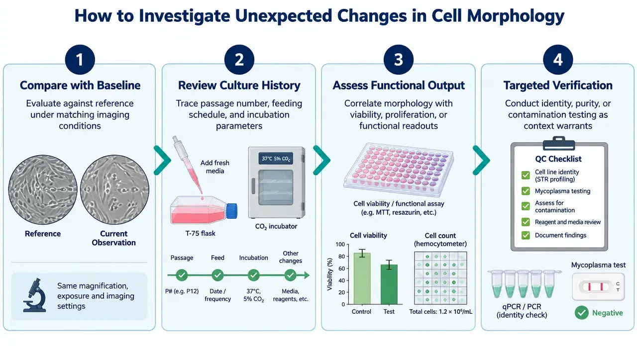
Use this four-step workflow to organize troubleshooting before changing culture conditions or interpreting experimental results.
1. Compare with a baseline
Review reference images from the same cell line under comparable conditions. Record passage number, seeding density, confluence, and observation time.
2. Review recent culture events
Check for changes in medium, serum, reagents, culture vessels, passaging, incubation conditions, or experimental treatments.
3. Assess functional changes
Determine whether morphology changes alongside cell number, proliferation, viability, or a phenotype relevant to the study.
4. Verify when necessary
Use appropriate contamination tests, cell-line authentication, or targeted assays when the evidence suggests a quality or biological issue.
Practical tip: If several cultures change at the same time, investigate shared reagents, equipment, and culture conditions. If the change occurs in only one culture, examine its individual handling history and contamination risk. Neither pattern proves a particular cause, but both help prioritize the investigation.
When Does Morphology Affect Experimental Results?
Morphological changes are particularly important when they influence the endpoint being measured or the interpretation of the data.
Cell Migration Assays
Changes in spreading, adhesion, and proliferation can influence scratch closure. Consider initial cell coverage, cell viability, and proliferation before attributing differences in closure rate to migration alone.
Drug Screening and Cytotoxicity
Rounding and detachment can indicate a cellular response but cannot independently establish apoptosis, necrosis, or another mechanism. Combine morphology with appropriate viability or cell-death assays.
Differentiation and Phenotypic Studies
Shape changes may accompany differentiation, but morphology alone does not confirm a specific cell identity or functional state. Use appropriate molecular markers and, where relevant, functional measurements.
Cell Model Reproducibility
Differences between culture batches may affect experimental consistency. Standardized imaging, culture records, cell sourcing, and relevant quality-control checks help distinguish normal variation from potential model problems.
Can Cell Morphology Be Quantified?
When visual inspection is insufficient, quantitative image analysis can provide more objective comparisons between cells, treatments, or experimental batches.
Cell Area
Measures the projected area occupied by a cell. It can help compare spreading or cell size under standardized imaging conditions.
Aspect Ratio
Describes relative elongation based on the cell's major and minor dimensions.
Circularity
Describes how closely a cell's outline resembles a circle. It does not identify the biological cause of rounding.
Reliable comparisons require consistent image acquisition and segmentation. Quantitative morphology describes a phenotype; additional evidence is needed to explain its cause.
Need to Evaluate Your Cell Model?
Reliable cell-based experiments start with appropriate model selection and quality control. Explore relevant cell characterization, contamination testing, or cell-based assay services to support your research needs.
References
- Ladke VS, Kumbhar GM, et al. Isolation, Culture and Morphological Assessment of Primary Cell Lines from Human Primary Oral Squamous Cell Carcinoma Using Explant Technique. Asian Pac J Cancer Prev. 2023 Jan 1;24(1):257-266.
- Lopes B R P, Ribeiro A G, et al. Diagnosis and treatment of HEp-2 cells contaminated with mycoplasma. Brazilian Journal of Biology. 2021 Feb 81(1):37-43
- Menicacci B, Cipriani C, et al, Giovannelli L. Modulation of the Senescence-Associated Inflammatory Phenotype in Human Fibroblasts by Olive Phenols. Int J Mol Sci. 2017 Oct 30;18(11):2275.
- Vargas-Accarino E, Herrera-Montávez C, et al. Spontaneous Cell Detachment and Reattachment in Cancer Cell Lines: An In Vitro Model of Metastasis and Malignancy. Int. J. Mol. Sci. 2021, 22(9), 4929