
How to Improve ISH Signal-to-Noise Ratio
In situ hybridization (ISH) allows researchers to visualize specific DNA or RNA sequences directly within cells and tissues. However, a successful experiment is not simply one that produces a strong signal. The target must be clearly distinguishable from background staining, with its location and distribution remaining biologically meaningful.
If your ISH images show weak staining, diffuse background, or inconsistent signals across tissue sections, the problem may lie in probe design, sample preparation, hybridization, washing, or detection. Understanding how these factors interact can help you troubleshoot more efficiently and improve signal-to-noise ratio (SNR) without compromising specificity.
What Causes a Poor ISH Signal-to-Noise Ratio?
Two problems commonly affect ISH results: the target signal is too weak, or the background signal is too strong.
Weak signals may result from low target abundance, degraded RNA, poor probe quality, or limited probe access to the target sequence. High background, on the other hand, may arise from nonspecific probe binding, inadequate washing, tissue autofluorescence, or inappropriate detection conditions.
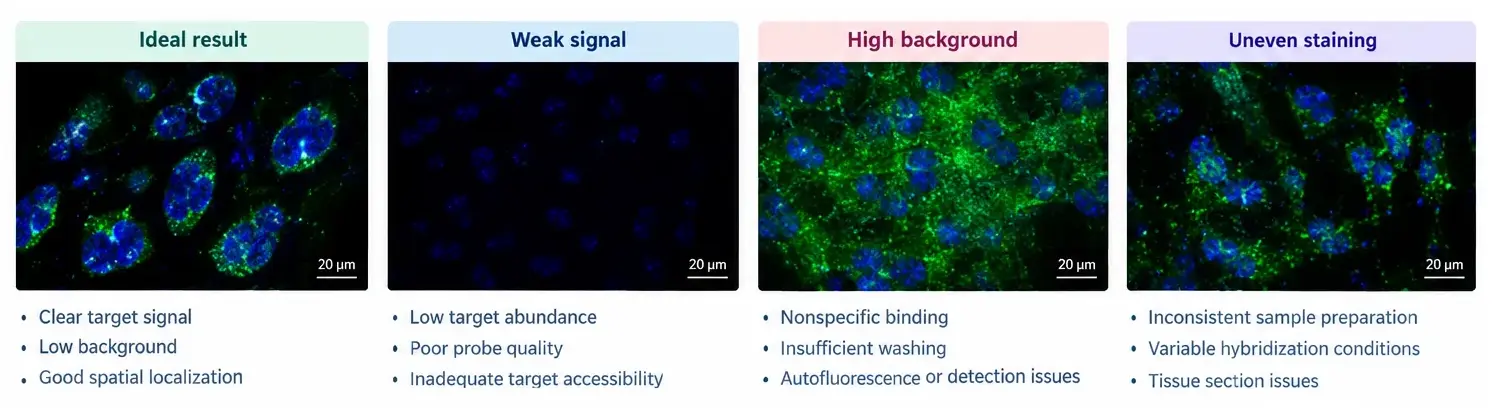
These problems can also occur together. For example, a sample that has been over-fixed may show weak target detection because the nucleic acid is less accessible to probes. Increasing probe concentration without addressing the underlying problem may then increase background without substantially improving the specific signal.
A useful starting point is to determine whether the main issue is target detection, background formation, or both. The following six strategies can help identify where optimization is most likely to make a difference.
1. Start with Probe Design and Quality
Even well-optimized experimental conditions cannot fully compensate for a poorly designed or degraded probe.
Probe specificity is particularly important when the target sequence shares similarities with other regions of the genome or transcriptome. Unintended binding can produce signals outside the expected target-positive cells or tissue regions. Probe length also matters: very short probes may have limited target-binding stability, while longer probes may penetrate some tissues less efficiently.
Before changing hybridization conditions, check that:
- The probe sequence matches the intended target and has acceptable specificity.
- Probe integrity, concentration, and labeling quality meet the requirements of the assay.
- The selected target region is suitable for the intended sample and detection method.
- Probe concentration is appropriate for the target abundance and assay chemistry.
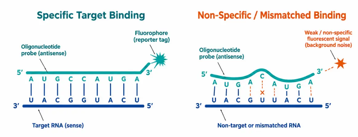
For example: If increasing probe concentration makes the entire tissue appear brighter but does not improve the distinction between target-positive and target-negative regions, the problem may not be insufficient probe input. Reducing the concentration or reassessing probe specificity may be more useful.
Probe design should therefore aim for specific, interpretable target detection rather than maximum signal intensity alone.
2. Optimize Sample Preparation Before Adjusting the Assay
Sample preparation is often an overlooked source of poor ISH performance. Fixation, permeabilization, and tissue processing affect both nucleic acid preservation and probe accessibility.
Insufficient fixation can compromise tissue integrity and allow RNA loss or degradation. Excessive fixation can make target sequences less accessible, sometimes requiring further optimization of pretreatment or digestion. These effects are well documented in tissue-based ISH methods.
To troubleshoot sample preparation:
- Confirm that fixation conditions are appropriate for the tissue type and assay.
- For paraffin-embedded sections, ensure that deparaffinization is complete.
- Optimize protease digestion or permeabilization to balance probe access with tissue preservation.
- Maintain consistent sample handling and prevent tissue sections from drying during the procedure.
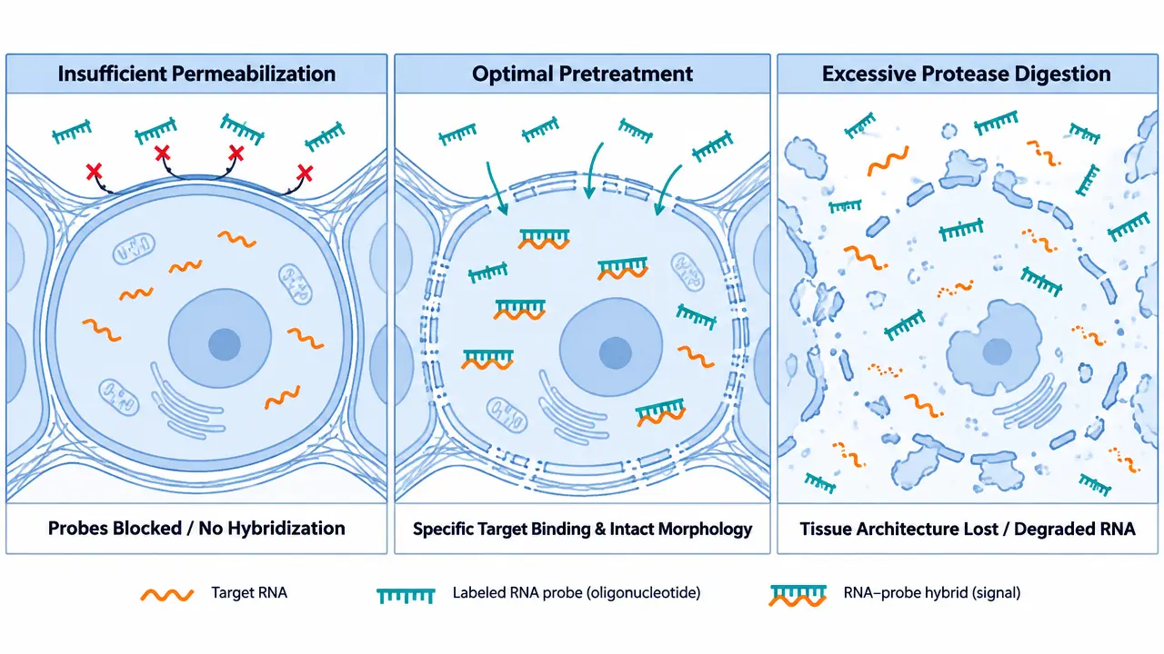
A practical example: Suppose two batches of paraffin-embedded tissue show different staining intensity despite using the same probe and hybridization conditions. Before changing the probe concentration, compare fixation history, tissue processing, and pretreatment conditions. Differences in these steps may explain the inconsistent results.
More digestion is not always better. Under-digestion can restrict probe access, whereas over-digestion can damage tissue morphology and contribute to abnormal staining. The appropriate balance depends on the sample and detection platform.
3. Find the Right Hybridization Conditions
Hybridization temperature, incubation time, buffer composition, and stringency all influence how effectively a probe binds its target.
Conditions that are insufficiently stringent may allow mismatched or nonspecific hybrids to remain. Conditions that are too stringent may weaken target binding and reduce the detectable signal.
If background is high, review whether the hybridization conditions adequately discriminate between complementary and non-complementary sequences. If the signal is weak, check whether the conditions are appropriate for the probe's sequence and binding characteristics.
A small, controlled optimization experiment is usually more informative than changing several parameters simultaneously. For example, compare a limited range of temperatures while keeping probe concentration, sample preparation, and washing conditions constant.
For microRNA ISH, probe-specific hybridization temperatures can be especially important. When multiple probes have different optimal temperatures, simultaneous hybridization may require a compromise, whereas sequential hybridization can allow each probe to be used under its preferred conditions.
The key is to identify conditions that preserve specific target binding while limiting unwanted interactions. There is no single hybridization temperature or incubation time that works for every ISH assay.
4. Use Post-Hybridization Washing to Remove Unwanted Signal
Washing helps remove unbound probes and destabilize nonspecific hybrids. If washing is insufficient, residual probes may contribute to diffuse background or staining in unexpected regions.
However, increasing wash stringency too aggressively can also remove weakly bound target-specific probes. The result may be a cleaner-looking image with less detectable target signal.
When troubleshooting, review wash-buffer composition, temperature, duration, and consistency between samples. Follow the recommendations for the specific probe and detection system, then make controlled adjustments if necessary.
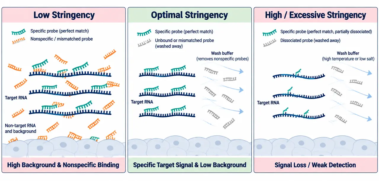
For example: If the target signal is clearly visible but diffuse staining also appears throughout the tissue, a carefully optimized increase in wash stringency may improve contrast. If the target signal disappears at the same time, the conditions may be too stringent, or the probe-target interaction may be insufficiently stable.
Technical reviews of RNA-FISH protocols identify post-hybridization washing as an important step for removing nonspecific hybrids and unbound probes. They also emphasize the need to balance background removal against signal retention.
5. Check Whether the Background Comes from Detection
Not all background originates during hybridization. The detection system, tissue properties, and imaging settings can also affect the final result.
For fluorescence ISH, tissue autofluorescence and spectral overlap between fluorophores can make it difficult to distinguish true target signals from unwanted fluorescence. For chromogenic ISH, nonspecific detection-reagent binding or excessive substrate development may contribute to background staining.
Consider the following checks:
For fluorescence ISH:
- Examine the relevant negative controls for autofluorescence.
- Check whether signals appear in unexpected channels.
- Verify fluorophore and filter compatibility.
- Keep exposure and acquisition settings consistent when comparing samples.
For chromogenic ISH:
- Review blocking and detection-reagent conditions.
- Check that washing steps are adequate.
- Avoid excessive substrate development that obscures signal localization.
A useful example comes from single-molecule FISH troubleshooting: fluorescence from one channel can bleed into another and create apparent spots that do not represent the intended target. Imaging each probe separately can help determine whether the problem originates from the detection setup rather than nonspecific hybridization.
6. Use Controls to Confirm That Optimization Worked
A brighter image does not necessarily indicate a better ISH result. The real objective is to improve the distinction between specific target signals and background while preserving meaningful spatial information.
Appropriate controls help determine whether the assay is working as intended:
- Positive controls help verify that the target can be detected under the selected conditions.
- Negative controls help identify nonspecific staining and background.
- RNA quality controls, where appropriate, help assess whether the sample and workflow support reliable RNA detection.
For example, if both the target sample and negative control become brighter after increasing probe concentration, the change may have increased background rather than improved specificity.
When possible, compare images using consistent acquisition settings and evaluate target intensity alongside background, localization, and tissue morphology. For quantitative studies, apply the same image-analysis criteria across samples.
The appropriate controls depend on the assay. A no-probe control can reveal some detection-related background, while a suitable negative probe or target-negative sample may provide additional information about nonspecific hybridization. No single control identifies every possible source of error.
ISH Troubleshooting: Common Problems and Solutions
These are starting points for troubleshooting, not universal diagnoses. Similar staining patterns can have different causes, so changes should be guided by the experimental evidence and the requirements of the specific ISH platform.
| Observed Problem | Possible Cause | What to Check First |
|---|---|---|
| Weak or absent signal | Low target abundance, degraded RNA, poor probe quality | Sample integrity, probe quality, positive controls |
| Diffuse background | Nonspecific binding, excessive probe concentration, insufficient washing | Probe specificity, concentration, wash conditions |
| Uneven staining | Inconsistent fixation, digestion, or hybridization | Sample preparation and incubation consistency |
| Signal lost after washing | Excessive wash stringency or unstable probe binding | Hybridization and washing conditions |
| High fluorescence background | Autofluorescence or channel bleed-through | Negative controls, fluorophore channels, imaging settings |
| Unexpected nuclear staining | Suboptimal pretreatment or tissue-specific effects | Platform-specific pretreatment and control results |
Need Help Optimizing Your ISH Assay?
Improving ISH signal-to-noise ratio often requires balancing probe specificity, sample preparation, hybridization conditions, and detection performance. Reviewing these factors systematically can help identify the source of poor signal quality and establish a more reproducible workflow.
Explore our related ISH services and technical resources to learn more about available assay options for your research needs.
Creative Bioarray Relevant Recommendations
| Products & Services | Description |
|---|---|
| Probe Development & Quality Control | Our Probe Development & Quality Control services focus on the end-to-end engineering and validation of FISH probes, from rational sequence design and synthesis to functional testing and performance verification. |
| ISH/FISH Analysis for Therapeutic R&D | High-resolution molecular and spatial analysis platforms designed to accelerate oncology and cell therapy research with clarity, sensitivity, and translational relevance. |