
How to Use iPSC-Derived Hepatocytes in Toxicity Studies
Drug-induced liver injury (DILI) remains a major challenge in drug development, particularly because hepatotoxicity can involve multiple cellular processes, including metabolic dysfunction, mitochondrial injury, oxidative stress, steatosis, and transporter-related effects. Traditional in vitro models provide useful information, but their ability to maintain human hepatic function over extended culture periods can vary.
iPSC-derived hepatocytes (iPSC-Heps) provide a renewable human cell model for investigating these responses. They can be used for early compound screening, mechanistic toxicology, drug metabolism studies, and longer-term exposure experiments. However, the value of an iPSC-derived hepatocyte model depends heavily on how the cells are qualified, how exposure is designed, and which endpoints are measured.
This guide outlines the key considerations for using iPSC-derived hepatocytes in toxicity studies, from model selection through data interpretation.
What are iPSC-Derived Hepatocytes?
iPSC-derived hepatocytes are generated by differentiating induced pluripotent stem cells toward the hepatic lineage. Depending on the differentiation and culture system, these cells can exhibit key hepatocyte functions, including albumin production, urea synthesis, CYP activity, and transporter function.
For toxicity studies, functional characteristics are more informative than marker expression alone. Cell maturity, metabolic activity, and baseline viability should therefore be considered when selecting a model.
When Should iPSC-Derived Hepatocytes Be Used?
iPSC-derived hepatocytes are particularly useful when toxicity studies require human-relevant hepatic responses, mechanistic characterization, or extended exposure.
Typical applications include:
- Early-stage compound screening
- DILI assessment
- Drug metabolism-related toxicity
- Mitochondrial and oxidative stress studies
- Steatosis and lipid-related toxicity
- Repeated-dose or longer-term exposure
- Mechanism-focused hepatotoxicity studies
The appropriate culture format and endpoint panel should then be selected according to the specific toxicity question.
How to Design an iPSC-Derived Hepatocyte Toxicity Study
A useful toxicity study starts with the biological question rather than the assay.
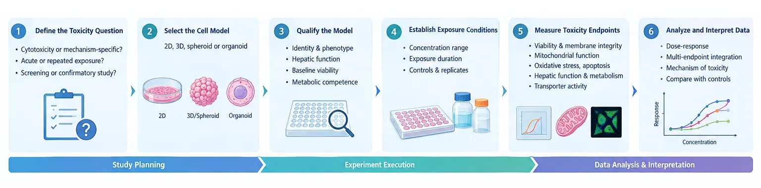 Fig. 1. Workflow for an iPSC-derived hepatocyte toxicity study.
1. Define the Toxicity Question
Before selecting endpoints, determine what the study needs to establish:
- General cytotoxicity or mechanism-specific toxicity?
- Acute or repeated exposure?
- Compound screening or confirmatory testing?
- Metabolic toxicity, mitochondrial injury, steatosis, or transporter-related effects?
- Direct cellular toxicity or toxicity associated with drug metabolism?
The answers determine the appropriate cell model, exposure window, concentration range, and analytical endpoints.
2. Select the Appropriate Cell Model
The choice between conventional 2D cultures, 3D cultures, spheroids, and other advanced hepatic models should reflect the study objective.
A 2D iPSC-hepatocyte model may be suitable for relatively high-throughput screening and straightforward imaging-based assays. Three-dimensional systems may be more useful when longer culture periods, cell-cell interactions, or repeated exposure are important.
The choice should also consider the functional characteristics of the specific iPSC-hepatocyte product or cell line. Relevant information includes differentiation consistency, baseline viability, hepatic function, CYP activity, and transporter activity.
3. Qualify the Model Before Compound Exposure
Baseline characterization is essential for interpreting toxicity data.
Useful qualification parameters include:
Identity and phenotype: hepatocyte-associated markers and morphology.
Hepatic function: albumin production, urea synthesis, and relevant CYP or transporter activities.
Baseline viability: consistent cell health before compound treatment.
Metabolic competence: where drug metabolism is central to the study, the activity of relevant enzymes should be established before interpreting compound responses.
This step helps distinguish a compound-specific effect from an underlying limitation of the cell model.
4. Establish the Exposure Design
Concentration and exposure duration should be selected with both biological relevance and assay performance in mind.
A concentration-response design is generally more informative than testing a single concentration. The exposure window should also reflect the toxicity question. Short exposure may capture acute cellular injury, whereas repeated or extended exposure can reveal changes in metabolism, mitochondrial function, lipid handling, or other processes that develop over time.
Vehicle controls, untreated controls, and appropriate positive controls should be included. Replicates and plate-level controls are also important for distinguishing biological effects from assay variability.
Key Toxicity Endpoints for iPSC-Derived Hepatocytes
No single endpoint provides a complete picture of hepatotoxicity. A combination of complementary measurements can provide more useful information.
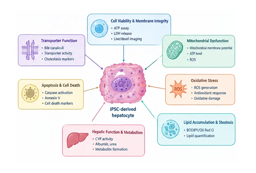 Fig. 2. Key toxicity endpoints in iPSC-derived hepatocyte.
Cell Viability and Membrane Integrity
Viability assays are often used as the first measure of overt toxicity. ATP-based assays, membrane integrity measurements, LDH release, and live/dead imaging can identify compounds that cause substantial cellular injury.
However, reduced viability alone does not explain the mechanism of toxicity. It is therefore best used as one component of a broader endpoint panel.
Mitochondrial Dysfunction
Mitochondrial effects are relevant to several forms of drug-induced liver injury. Depending on the study design, measurements may include ATP levels, mitochondrial membrane potential, mitochondrial mass, and reactive oxygen species (ROS).
A compound that produces mitochondrial dysfunction before substantial loss of viability may warrant further mechanistic investigation.
Apoptosis and Cell Death
Apoptosis-related assays can help distinguish programmed cell death from general loss of viability. Caspase activation, Annexin V staining, and other cell-death markers can be combined with viability measurements to establish a clearer response profile.
Oxidative Stress
ROS generation and related stress responses can provide additional information when oxidative damage is suspected. These measurements are particularly useful when interpreted alongside mitochondrial and cell-death endpoints rather than as isolated results.
Lipid Accumulation and Steatosis
Changes in intracellular lipid content can be used to investigate drug-induced steatosis and disruption of hepatic lipid metabolism. Imaging-based lipid measurements can also be combined with viability and functional assays to determine whether lipid accumulation occurs before overt cellular injury.
Hepatic Function and Drug Metabolism
Functional endpoints are particularly valuable when the study involves compounds that may alter hepatic metabolism.
Depending on the objective, analysis may include:
- CYP enzyme activity
- metabolite formation
- albumin production
- urea synthesis
- phase II metabolic activity
- transporter function
A reduction in metabolic activity should not automatically be interpreted as cell death. It may reflect altered hepatic function, enzyme inhibition, cellular stress, or changes in differentiation state.
Bile Canalicular and Transporter Function
Transporter-related effects are relevant to cholestatic mechanisms of liver injury. Assessment of bile canalicular structures or transporter activity can provide information that is not captured by conventional viability assays.
2D vs 3D iPSC-Derived Hepatocytes
The choice between 2D and 3D culture depends largely on the study requirements.
| Feature | 2D Culture | 3D Culture |
|---|---|---|
| Throughput | Generally higher | Generally lower |
| Experimental complexity | Lower | Higher |
| Cell-cell interactions | More limited | More extensive |
| Long-term studies | Application-dependent | Often advantageous |
| Imaging | Straightforward | More complex |
For early compound screening, 2D systems can offer simpler handling and higher throughput. For longer studies, 3D cultures may provide a more suitable environment for maintaining cellular organization and hepatic function.
Neither format should be considered universally superior. The relevant question is whether the model provides the functional characteristics needed for the intended toxicity endpoint.
iPSC-Derived Hepatocytes vs PHHs vs HepG2
Different liver cell models serve different purposes.
| Model | Key Strength | Main Consideration | Typical Application |
|---|---|---|---|
| HepG2 | Easy to culture and scalable | Limited hepatic metabolic function | Early cytotoxicity screening |
| PHHs | Strong human hepatic functionality | Donor variability and limited availability | Drug metabolism and hepatotoxicity |
| iPSC-derived hepatocytes | Renewable human cell source | Maturity and functional variability | Toxicity and mechanistic studies |
| 3D iPSC-hepatocytes | Suitable for extended culture | More complex assay workflow | Long-term and repeated exposure |
Rather than selecting a single model for every stage, complementary models can be used to answer different questions during drug development.
Can iPSC-Derived Hepatocytes Predict DILI?
iPSC-derived hepatocytes can provide useful information for DILI-related screening and mechanistic studies, particularly when multiple toxicity pathways are evaluated together.
They can capture responses associated with cellular injury, mitochondrial dysfunction, oxidative stress, steatosis, metabolic disruption, and transporter effects. However, predictive performance can vary with hepatocyte maturity, differentiation protocol, culture format, compound characteristics, exposure conditions, and endpoint selection.
For this reason, iPSC-derived hepatocytes are best viewed as part of a broader toxicity assessment strategy rather than a standalone predictor of clinical liver injury.
How to Interpret Toxicity Data
Data interpretation should consider both the magnitude of the response and the relationship between different endpoints.
For example, a reduction in viability accompanied by increased ROS, loss of mitochondrial membrane potential, and activation of apoptosis markers provides a stronger indication of a defined toxicity process than a viability change alone.
Concentration-response relationships are also important. A response observed only at concentrations far above relevant exposure levels may have different implications from a response occurring within a biologically relevant range.
Functional changes should likewise be interpreted carefully. Reduced albumin production, CYP activity, or transporter function may indicate impaired hepatic function without necessarily representing irreversible cell death.
Common Challenges and Experimental Considerations
Several factors can influence the reliability of iPSC-derived hepatocyte toxicity data:
- Cell-line variability: different iPSC lines may exhibit different differentiation and functional profiles.
- Hepatocyte maturity: metabolic and transporter activities may not fully match those of adult primary hepatocytes.
- Batch variability: differences between differentiation or production batches should be monitored.
- Culture conditions: media, extracellular matrix, cell density, and culture format can affect hepatic function.
- Assay interference: compounds may interfere with fluorescence, luminescence, or other assay signals.
- Endpoint selection: relying on a single readout can miss important toxicity mechanisms.
These factors should be considered during study planning rather than addressed only after unexpected results appear.
Creative Bioarray Relevant Recommendations
| Products & Services | Description |
|---|---|
| Human iPSC-derived Hepatocytes | iPSC-derived hepatocytes are developed to overcome deficiencies found in traditional methods and systems. Human iPSC-derived Hepatocytes express biomarkers (e.g., E-Cadherin and albumin). |