
Why Is My IHC Background Too High? A Step-by-Step Troubleshooting Guide
High background staining is one of the most common—and frustrating—problems in immunohistochemistry (IHC). Instead of seeing a clear, specific signal in the expected cells or tissue compartment, you may observe diffuse brown staining, widespread haze, dark tissue edges, granular deposits, or staining in your negative controls.
When IHC background is too high, the result is not simply "ugly." Excessive background can mask weak target expression, reduce confidence in scoring, create false-positive interpretations, and make image analysis unreliable.
The good news is that high IHC background is usually diagnosable. The key is to avoid changing every parameter at once. By examining the staining pattern, using the right controls, and optimizing variables in a logical order, you can identify whether the issue comes from the primary antibody, secondary detection system, blocking step, antigen retrieval, endogenous enzyme activity, or chromogen development.
This guide explains how to troubleshoot high IHC background step by step.
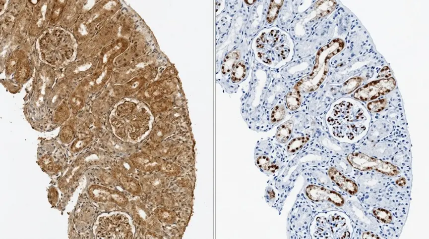 Fig. 2. High IHC background versus optimized target staining. Excessive diffuse DAB staining
can obscure tissue morphology and reduce the signal-to-noise ratio, whereas an optimized protocol produces
localized, interpretable target staining with minimal non-specific background.
First: Is It Really Background?
Not every dark area is non-specific IHC staining. What looks like background may be:
- Genuine low-level expression of the target;
- Endogenous peroxidase activity;
- Endogenous biotin;
- Melanin, lipofuscin, hemosiderin, or blood-related pigment;
- Necrotic tissue staining;
- DAB precipitate;
- Tissue folds, damaged edges, or drying artifacts;
- Antibody cross-reactivity.
Ask whether the signal is located where your target should be. A membrane protein should not produce widespread nuclear staining. A nuclear transcription factor should not show only diffuse cytoplasmic signal. If the staining location does not match target biology, treat the result cautiously.
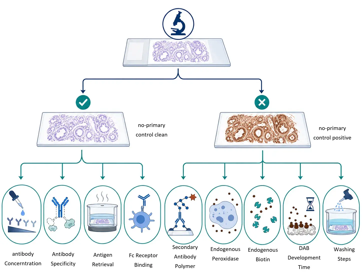 Fig. 2. Using a no-primary antibody control to identify the source of IHC background. A clean
no-primary control suggests that the primary antibody, antigen retrieval, or non-specific tissue binding may be
responsible. Persistent staining in the no-primary control points instead to the detection system, endogenous
enzymes, endogenous biotin, insufficient washing, or chromogen-related artifacts.
Start With the No-Primary Antibody Control
The no-primary control follows the full protocol but omits the primary antibody. It is one of the fastest ways to identify the source of high background.
If the no-primary control is clean
The problem is more likely related to:
- Primary antibody concentration;
- Primary antibody specificity;
- Antibody incubation time;
- Inadequate protein blocking;
- Fc receptor-mediated binding;
- Overly harsh antigen retrieval.
If the no-primary control is also positive
The background is likely caused by:
- Secondary antibody or polymer detection reagents;
- Endogenous peroxidase;
- Endogenous biotin;
- Excessive DAB development;
- Insufficient washing;
- Tissue pigment or another non-immunologic artifact.
This distinction prevents unnecessary antibody optimization when the real problem is the detection system.
Read the Staining Pattern Before Changing the Protocol
| Staining pattern | Common causes | First troubleshooting step |
|---|---|---|
| Uniform staining across the section | Antibody too concentrated, DAB overdevelopment, poor washing | Dilute antibody, shorten DAB, increase washes |
| Strong signal in blood-rich regions | Endogenous peroxidase | Optimize hydrogen peroxide blocking |
| Background in lymph node, spleen, or inflamed tissue | Fc receptor binding | Add Fc block or use F(ab')₂-based reagents |
| Dark section edges | Tissue drying, uneven reagent coverage | Keep slides wet and use sufficient reagent volume |
| Brown granular particles | DAB precipitate, contaminated reagents | Use fresh filtered DAB and clean containers |
| Unexpected nuclear staining | Cross-reactivity, harsh retrieval, antibody too concentrated | Titrate antibody and reduce retrieval strength |
| Signal in no-primary control | Detection system or endogenous tissue activity | Check peroxidase/biotin block and polymer system |
1. Primary Antibody Concentration Is Too High
An over-concentrated primary antibody may bind weakly to unrelated proteins or tissue components. This is one of the most common causes of diffuse IHC background.
You may suspect this issue if:
- The no-primary control is clean;
- Background decreases after antibody dilution;
- Specific staining remains visible at lower antibody concentration;
- Staining appears in biologically unexpected regions.
Instead of making one random adjustment, perform a titration. If your current dilution is 1:100, test 1:200, 1:400, and 1:800 using serial sections from the same tissue block.
Choose the condition with the best signal-to-background ratio—not simply the darkest staining.
You can also reduce the primary antibody incubation time. Overnight incubation may improve sensitivity, but it can increase background if the antibody concentration is already too high.
2. Secondary Antibody or Detection Polymer Is Too Strong
Highly sensitive HRP polymer systems can amplify weak target signals, but they can also amplify low-level non-specific binding.
This is particularly common when:
- The tissue contains endogenous immunoglobulins;
- The secondary antibody is not sufficiently cross-adsorbed;
- The secondary or polymer reagent incubation is too long;
- A highly sensitive detection system is paired with a strong primary antibody.
If the no-primary control is positive, reduce secondary/polymer incubation time, evaluate your blocking steps, and confirm that the detection reagent is appropriate for your primary antibody species.
For mouse primary antibodies used on mouse tissue, use a dedicated mouse-on-mouse detection system when possible. Conventional anti-mouse secondary antibodies can recognize endogenous mouse immunoglobulins and create strong background.
3. Endogenous Peroxidase Was Not Fully Blocked
In HRP-DAB IHC, endogenous peroxidase can produce unwanted brown staining. It is especially relevant in blood-rich, inflamed, or myeloid-cell-rich tissues.
Common high-risk tissues include:
- Spleen;
- Bone marrow;
- Liver;
- Kidney;
- Hemorrhagic tumors;
- Inflamed tissue;
- Samples with abundant red blood cells.
Typical signs include brown staining in erythrocyte-rich regions or persistent staining in no-primary controls.
Use an appropriate hydrogen peroxide blocking step before applying the primary antibody. If background persists, compare your standard block with a longer incubation, a fresh blocking solution, or a validated commercial peroxidase-blocking reagent.
Avoid making the block unnecessarily harsh, since over-treatment can damage tissue morphology or reduce antigen detection.
4. Endogenous Biotin Is Causing False Signal
If you use an avidin-biotin or streptavidin-biotin detection system, endogenous biotin can produce high background.
This is common in tissues such as liver, kidney, intestine, adrenal gland, brain, and mammary tissue.
If your no-primary control remains positive despite adequate peroxidase blocking, endogenous biotin may be responsible.
Possible solutions include:
- Adding an avidin/biotin blocking step;
- Using a biotin-free polymer detection system;
- Comparing results from ABC and polymer-based detection methods.
For many challenging tissues, biotin-free polymer detection is the simpler and more reliable option.
5. Protein Blocking or Fc Receptor Blocking Is Inadequate
Protein blocking reduces non-specific binding to tissue proteins and exposed cellular structures. Depending on your protocol, you may use normal serum, BSA, casein, or a commercial blocking reagent.
However, standard protein blocking may not be enough for immune-rich tissue.
Fc receptor-mediated background is common in:
- Lymph nodes;
- Spleen;
- Tonsil;
- Bone marrow;
- Inflamed tissue;
- Immune-cell-rich tumors.
If background is concentrated in macrophages, monocytes, or inflammatory infiltrates, add an Fc receptor blocking step. You may also consider highly cross-adsorbed secondary antibodies or F(ab')₂ antibody fragments to reduce Fc-associated binding.
6. Antigen Retrieval Is Too Harsh
Antigen retrieval is necessary for many FFPE samples, but excessive heat, high pH, or overly long retrieval can increase non-specific staining.
Signs of over-retrieval include:
- Damaged tissue morphology;
- Tissue detachment;
- Increased diffuse or nuclear staining;
- More background with pH 9 retrieval than pH 6 retrieval;
- Unexpected staining patterns.
Test a small retrieval matrix rather than relying on one standard condition:
| Condition | Example |
|---|---|
| Mild | Citrate buffer, pH 6, shorter retrieval |
| Standard | Citrate buffer, pH 6, standard retrieval |
| Strong | EDTA or Tris-EDTA, pH 9 |
| Reduced strong | pH 9 buffer with shorter retrieval |
Select the condition that preserves tissue structure and provides specific staining with the lowest background.
7. Washing Is Not Adequate
Insufficient washing leaves unbound antibodies and detection reagents on the tissue. This can increase diffuse background and produce inconsistent staining.
Review:
- Number of washes;
- Wash duration;
- Buffer volume;
- Gentle agitation;
- Slide spacing during washing;
- Buffer freshness;
- Whether a mild detergent is suitable for your assay.
Never allow sections to dry during the staining process. Drying can create dark edges, uneven staining, and irreversible artifacts.
8. DAB Development Time Is Too Long
DAB is sensitive and easy to overdevelop. Excessive development creates a muddy brown appearance, obscures morphology, and makes weak non-specific signal appear positive.
Test several DAB development times, such as 30 seconds, 1 minute, 2 minutes, and 5 minutes. Use the shortest time that produces a clear, interpretable target signal.
If you see granular brown particles, prepare DAB fresh, filter it if needed, and check for contaminated staining dishes or reagent containers.
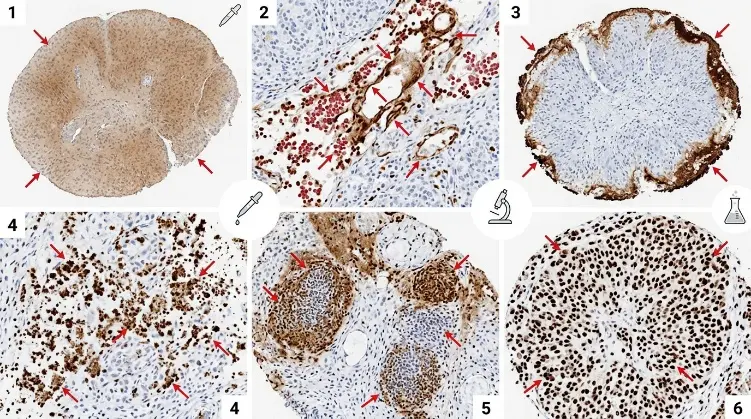 Fig. 3. Common high-background staining patterns in immunohistochemistry. Diffuse haze,
blood-associated staining, edge effects, granular precipitates, immune-cell-associated background, and
unexpected nuclear staining may arise from different technical causes and should be investigated using
pattern-specific troubleshooting steps.
A Six-Slide IHC Troubleshooting Test
When tissue is limited, use serial sections to test the problem efficiently.
| Slide | Condition | Purpose |
|---|---|---|
| 1 | Standard protocol | Baseline result |
| 2 | No-primary control | Identifies detection/endogenous background |
| 3 | Lower primary antibody concentration | Tests antibody overload |
| 4 | Shorter DAB development | Tests chromogen overdevelopment |
| 5 | Stronger peroxidase block | Tests endogenous peroxidase |
| 6 | Alternative protein block or Fc block | Tests non-specific/Fc binding |
How to interpret the result
- No-primary control is positive: Focus on endogenous enzymes, biotin, secondary/polymer reagents, washing, and DAB.
- Lower primary antibody improves the result: Optimize antibody dilution and incubation time.
- Shorter DAB improves the result: Your chromogen development is too long.
- Stronger peroxidase block improves blood-rich areas: Endogenous peroxidase is a major contributor.
- Fc block improves immune-rich tissue: Fc receptor binding is likely involved.
- None of these changes work: Reassess antibody specificity, clone selection, tissue preparation, and antigen retrieval.
When Should You Change the Antibody?
Protocol optimization cannot compensate for a poorly validated or cross-reactive antibody.
Consider switching antibodies or validating with a second clone if:
- Several antibody dilutions still produce high background;
- Signal localization does not match known biology;
- Different retrieval conditions do not improve specificity;
- Background occurs across unrelated tissue types;
- A second antibody against the same target produces a different pattern;
- Results are not supported by positive controls, RNA data, Western blotting, or another orthogonal method.
Final Takeaway
High IHC background is not one single problem. It can come from antibodies, tissue properties, blocking steps, detection chemistry, or chromogen development.
Start with the no-primary control, inspect the staining pattern, and adjust one major variable at a time. In many cases, a structured workflow—antibody titration, improved blocking, optimized antigen retrieval, stronger washing, and shorter DAB development—will produce a cleaner and more reliable IHC result.
Creative Bioarray Relevant Recommendations
| Products & Services | Description |
|---|---|
| Immunohistochemistry (IHC), Immunofluorescence (IF) Service | Creative Bioarray offers a comprehensive IHC service from project design, marker selection to image completion and data analysis. |
| Histology Services | Creative Bioarray offers tissue processing, embedding, sectioning, and staining, along with set of histological examination services. |