
Cell Clumping in Suspension Culture: Causes & Solutions
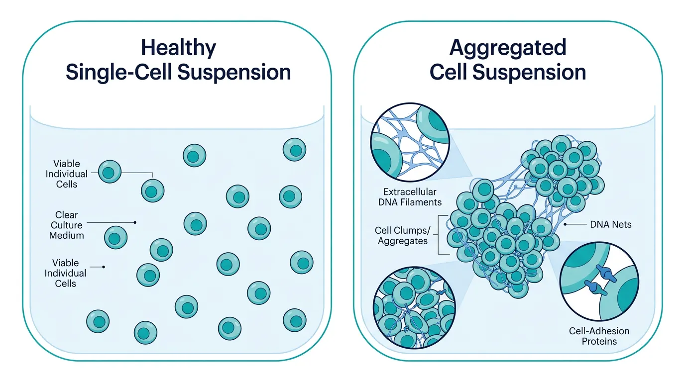
Managing suspension cultures—whether for biomanufacturing in CHO and HEK293 lines, T-cell expansion, or stem cell aggregates—requires a delicate balance of physical and chemical parameters. Uncontrolled cell clumping is one of the most persistent bottlenecks in bioprocessing. Beyond making automated counting and sampling unreliable, dense cell aggregates limit nutrient and oxygen diffusion, trigger localized necrosis, and block downstream filtration systems.
Resolving cell aggregation begins with identifying the underlying physical, chemical, or biological drivers behind the clumps.
Quick Diagnostic Flowchart
When cell aggregates form in your suspension vessels, match the physical appearance of the clumps to these three primary diagnostic paths:
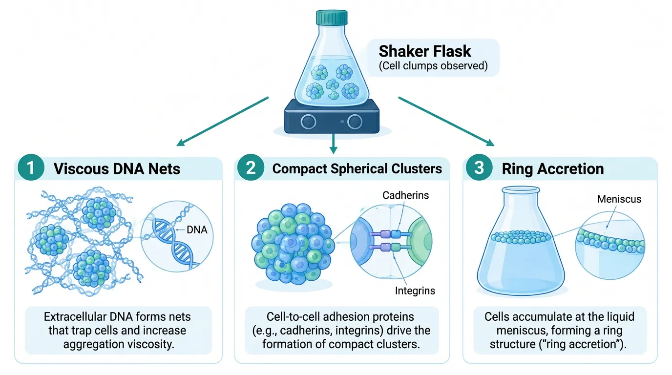
Pathway A: Viscous, Thread-Like Aggregates
- What you see: Irregular, sticky clumps that string together like a web and resist gentle dissociation.
- Likely culprits: Cell lysis, genomic DNA release, and high dead-cell ratios creating extracellular DNA nets.
Pathway B: Compact, Spherical Clusters
- What you see: Smooth, tight cell spheres formed even when viability remains high (>90%).
- Likely culprits: Calcium/magnesium-dependent cell adhesion molecules (cadherins/integrins) or high cell density.
Pathway C: Ring-Like Aggregation or Wall Accretion
- What you see: Cell rings forming along the fluid meniscus or heavy accumulation at the bottom of the flask.
- Likely culprits: Suboptimal shaking speeds, incorrect orbital stroke diameters, or improper vessel geometry.
The Mechanisms Driving Cell Aggregation
To stop clumping effectively, it helps to look at the forces binding these cells together.
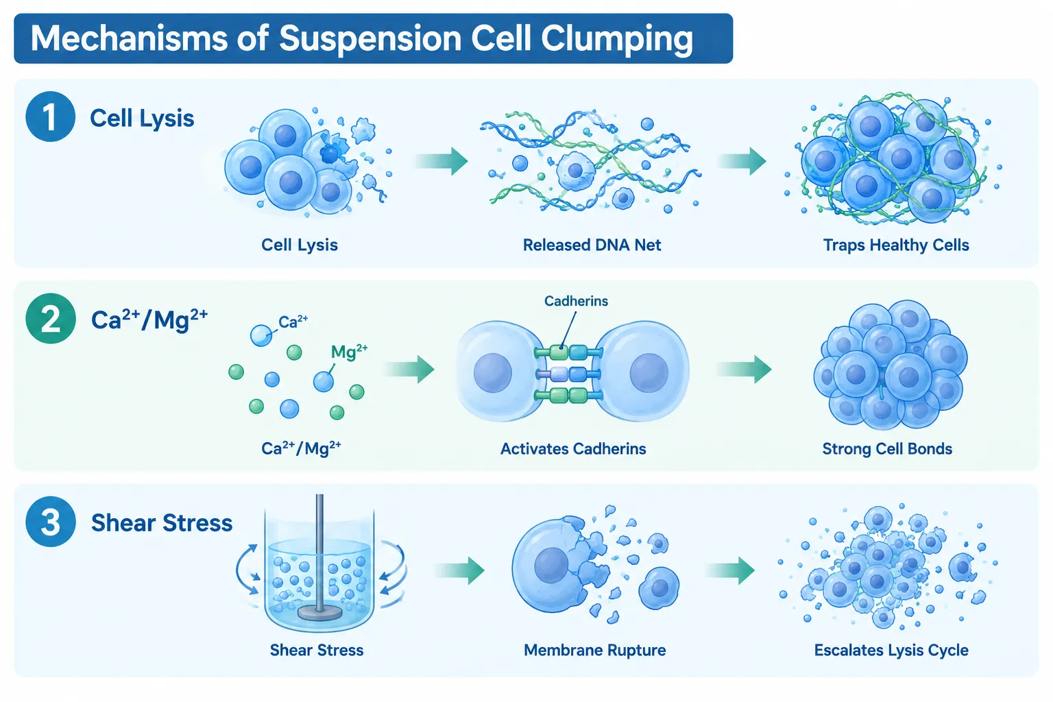
Extracellular DNA (eDNA) and "DNA Nets"
When suspension cells lyse—whether due to overconfluency, rapid thawing, or shear stress—they release long, sticky strands of high-molecular-weight genomic DNA into the media. This extracellular DNA acts like a microscopic net. As the culture shakes, these sticky strands sweep through the suspension, entangling healthy live cells and forming large, sticky aggregates.
Divalent Cation-Mediated Adhesion
Cell-surface adhesion proteins, such as integrins and cadherins, require divalent cations (Ca2+ and Mg2+) to maintain their active binding conformations. In media containing high concentrations of calcium or magnesium, or when specific recombinant lines overexpress surface adhesion markers, cells naturally stick to one another upon physical collision.
Hydrodynamic Forces and Shear Stress
Suspension cultures rely on fluid movement for gas exchange and mass transfer. If the agitation speed is too low, gravity takes over, allowing local cell densities to spike and form clusters. Conversely, if the agitation speed is too high or the impeller creates excessive shear stress, cells rupture. This unleashes more eDNA and dead-cell debris, accelerating the clumping cycle.
Root Causes & Step-by-Step Solutions
Issue 1: High Lysis Rates and Viscous Clumping
The Cause: Culture viability drops below 85–90%, or cells were subjected to harsh thawing protocols, releasing genomic DNA.
The Fix: Supplement the culture media with DNase I (20–50 μg/mL) or enzymatic dissociation reagents like Accutase to digest the extracellular DNA strands without damaging intact cell membranes. Maintain subculture schedules to harvest or passage cells before they reach late stationary phase.
Issue 2: Spontaneous Aggregation in High-Viability Cultures
The Cause: Media formulations rich in calcium or magnesium trigger cell-surface cadherin interaction.
The Fix: Transition your culture to low-calcium/magnesium chemically defined media formulations. Alternatively, introduce commercial non-ionic anti-clumping agents or surfactants such as Pluronic F-68 (0.1% w/v) or dextran sulfate. These additives lower surface tension and sterically block cell-to-cell adhesion.
Issue 3: Inadequate Hydrodynamics and Vessel Sedimentation
The Cause: A mismatch between the orbital shaker diameter, flask shape, and fluid volume causes poor fluid mixing or excessive shear.
The Fix: Adjust agitation speeds based on your shaker’s throw (orbital diameter). Switch from plain-bottom flasks to baffled flasks if you need higher oxygen transfer without settling, or move to plain-bottom flasks if baffled designs are causing shear-induced cell lysis.
Tailored Anti-Clumping Protocols
Different cell platforms require specialized strategies to maintain single-cell suspensions:
| Cell Platform | Representative Models | Primary Cause of Clumping | Recommended Anti-Clumping Strategy | Operational Pro-Tip |
|---|---|---|---|---|
| Industrial Production | CHO-K1, High-Yield HEK293 | High cell density & membrane debris | Dextran sulfate or commercial anti-clumping supplements | Add anti-clumping agents early during seed-train adaptation; remove before transfection if required. |
| Immune & T-Cells | Jurkat, Primary CAR-T Cells | Activation-induced surface marker clustering | Low-binding surface vessels + Recombinant DNase | Keep agitation gentle; avoid high shear during expansion phases. |
| Stem Cell Spheres | iPSCs, Neural Progenitors | Cation-dependent cadherin adhesion | Selective Y-27632 (ROCK inhibitor) + Accutase | Dissociate to single cells using gentle enzymatic steps; re-seed at controlled densities. |
| Primary Suspensions | Splenocytes, PBMC Fractions | Sticky eDNA from isolation steps | DNase I treatment during washing | Filter suspension through a 40 $\mu\text{m}$ cell strainer prior to counting or plating. |
Golden Rules to Prevent Cell Clumping
- Maintain High Viability (>90%): The best defense against clumping is preventing cell lysis. Passage cultures while they are still in the exponential growth phase.
- Add Anti-Clumping Supplements Proactively: Do not wait for massive aggregates to appear. Introduce surfactants or anti-clumping agents during the initial adaptation phase to suspension culture.
- Match Shaker Hydraulics to Vessel Size: Ensure the liquid volume stays between 20% and 30% of the total flask capacity for optimal mixing without extreme shear.
- Handle Suspensions Gently: Use wide-orifice pipette tips for mixing. Avoid aggressive vortexing or forced passaging through small-gauge needles.
- Audit for Contamination: Silent bacterial or fungal contamination often presents as sudden, unexplained cell clumping and media cloudiness.
Frequently Asked Questions
Q1: Can I use Trypsin to break up cell clumps in my suspension bioreactor?
Trypsin is generally not recommended for routine suspension cultures. It is a harsh, non-specific protease that degrades cell-surface receptors and membrane proteins, leading to lower viability over continuous passages. Instead, use recombinant enzymes, DNase I, or dedicated anti-clumping media additives.
Q2: Will anti-clumping agents affect my transient transfection efficiency?
Yes, some anti-clumping supplements (especially dextran sulfate or certain polyanionic additives) can interfere with lipid- or PEI-mediated transfection by binding to positive charges. It is best to omit anti-clumping agents 1–2 passages prior to transfection and add them back 24 hours post-transfection.
Q3: Why do cells clump heavily right after thawing from liquid nitrogen?
Thawing causes physical stress and cryoprotectant toxicity, leading to partial cell lysis. The released genomic DNA immediately forms sticky nets around surviving cells. To prevent this, wash thawed cells in warm media containing DNase I and centrifuge gently to discard released DNA before seeding.